- Remove All
 Your shopping cart is currently empty


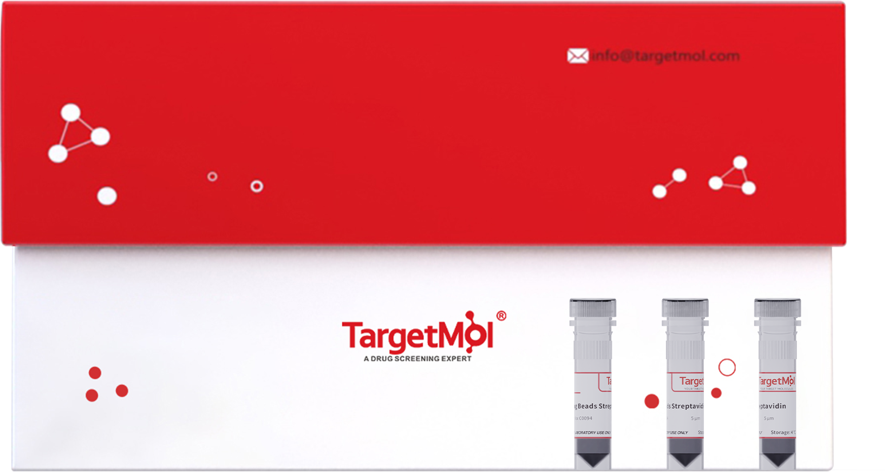
Mag Beads Streptavidin (5 μm)
TargetMol Mag Beads Streptavidin covalently connects SA to the beads surface through protein-protein coupling method. This enables efficient binding of biotinylated antibodies, nucleic acids, and proteins.
The product combines superparamagnetic advantages with unique features including uniform size and regular morphology, facilitating the rapid capture of target molecules and magnetic separation. This product can be used with automated equipment for high-throughput technology.
| Pack Size | Price | Availability | Quantity |
|---|---|---|---|
| 1 mL | $95 | In Stock | |
| 5 mL | $355 | In Stock | |
| 10 mL | $635 | In Stock |
 Advantages
- High binding capacity
- Good stability
- Quick and sensitive magnetic response
- Suitable for a wide range of experimental conditions
 Applications
- Immunoassays & Protein Isolation: Specifically binds to biotinylated antibodies or antigens, can be used as a solid-phase reaction carrier for immunoassays and ELISA.
- Nucleic acid isolation & Nucleic Acid Probe Preparation: Specifically binds to biotinylated nucleic acid probes, can be used for DNA and RNA hybridization experiments.
- DNA-protein interaction studies: Specifically binds to biotinylated target DNA or RNA fragments, can be used to study the interaction between proteins and nucleic acids.
- Cell isolation and activation: Can immobilize antibodies to specifically recognize target cells.
 Comparison of Streptavidin Magnetic Beads
| Product Name | C0089 | C0090 | C0091 | C0092 | C0093 | C0094 |
|---|---|---|---|---|---|---|
| Particle Size | 300 nm | 1 μm | 2 μm | 2.8 μm | 3 μm | 5 μm |
| Binding Capacity for Biotin-Nucleic Acid Probes (24nt) (pmol/mg beads) | ≥450 | ≥450 | ≥350 | ≥300 | ≥200 | ≥300 |
| Binding Capacity for Biotin-Rabbit IgG (μg/mg beads) | ≥15 | ≥15 | ≥15 | ≥10 | ≥5 | ≥10 |
| Applications | Cell Isolation; Nucleic acid probe capture | Immunoprecipitation; DNA-protein interaction | Immunoprecipitation; DNA-protein interaction | Magnetic particle-based chemiluminescence; Neutralizing antibody detection | Magnetic particle-based chemiluminescence | Cell activation |
| Bead Concentration | 10 mg/mL | 10 mg/mL | 10 mg/mL | 10 mg/mL | 10 mg/mL | 10 mg/mL |
| Magnetic Beads Surface | Hydrophilic groups | Hydrophilic groups | Hydrophilic groups | Hydrophilic groups | Hydrophilic groups | Hydrophilic groups |
 Handling Instruction
1. Recommended Buffers
Note: Adjust salt concentration and pH as needed during use.
(1) Buffer I (Nucleic acid applications): 10 mM Tris-HCl (pH 7.5), 1 mM EDTA, 1 M NaCl, 0.01%-0.1% Tween-20.
(2) Buffer II (Antibodies/protein applications): PBS, pH 7.4 (0.05% Tween-20), add 0.01%-0.1% BSA based on needs.
(3) Washing Buffer (for chemiluminescence): Prepare washing buffer as needed, and equilibrate to room temperature before use.
2. Immobilization of biotinylated nucleic acids
(1) Resuspend the magnetic beads in the vial (or vortex for 20 seconds). Transfer 100 μL of beads into a new tube and place the tube into a magnetic stand to collect the beads against the side of the tube (Hereinafter referred to as magnetic separation). Remove and discard the supernatant.
Note: It’s recommended to add biotinylated molecules at 1-2 times of beads.
(2) Add 1 mL Buffer I to the beads and vortex fully to resuspend the beads. Remove and discard the supernatant from magnetic separation.
Note: If the bead volume used in step (1) exceeds 1 mL, add an equal volume of Buffer I.
(3) Repeat step (2) once.
(4) Add 500 μL of biotinylated nucleic acids diluted with Buffer I (making the bead at a final concentration of 2 mg/mL) and vortex the tube fully to resuspend the beads. Rotate the tube for 30 minutes at room temperature.
(5) Perform magnetic separation and transfer the supernatant to a new centrifuge tube.
(6) Wash beads three times as step (2).
(7) Add an appropriate amount of low salt buffer to resuspend the beads according to the requirements of subsequent experiments. The biotinylated nucleic acid binding step is complete, and the beads can be used for subsequent operations.
(8) The amount of nucleic acid can be calculated by measuring the nucleic acid concentration before and after the reaction: (concentration before reaction - concentration after reaction) × reaction solution volume.
3. Immobilization of biotinylated antibodies/proteins
(1) Resuspend the magnetic beads in the vial (or vortex for 20 seconds). Transfer 100 μL of beads into a new tube and place the tube into a magnetic stand to collect the beads against the side of the tube (Hereinafter referred to as magnetic separation). Remove and discard the supernatant.
Note: It’s recommended to add biotinylated molecules at 1-2 times of beads.
(2) Add 1 mL Buffer II to the beads and vortex fully to resuspend the beads. Remove and discard the supernatant from magnetic separation.
Note: If the bead volume used in step (1) exceeds 1 mL, add an equal volume of Buffer II.
(3) Repeat step (2) once and wash beads three times.
(4) Add 1 mL of biotinylated antibodies/proteins diluted with Buffer II (making the bead at a final concentration of 1 mg/mL) and vortex the tube fully to resuspend the beads. Rotate the tube for 60 minutes at room temperature.
(5) Perform magnetic separation and transfer the supernatant to a new centrifuge tube.
(6) Wash beads five times as step (2).
(7) Binding is now complete. Resuspend the beads in Buffer II or a buffer suitable for downstream applications to a desired concentration according to experimental needs.
4. Magnetic particle-based chemiluminescence immunoassay
(1) Adjust to an appropriate concentration (recommended 0.2-0.8 mg/mL) and resuspend the magnetic beads in the vial (or vortex for 20 seconds). Transfer 50 μL of beads into a 96-well plate and perform magnetic separation. Discard the supernatant and remove the plate.
(2) Add 100 μL of biotinylated capture antibody to each well and vertex the tube fully to resuspend the beads. Incubate at 37°C for 15 minutes. Perform magnetic separation and discard the supernatant. Remove the 96-well plate.
(3) Add 200 μL of washing buffer to each well and vertex the tube fully to resuspend the beads. Perform magnetic separation and discard the supernatant. Repeat this step twice, and wash beads three times totally.
(4) Add 100 μL of standards or test samples to each well and vertex the tube fully to resuspend the beads. Incubate at 37°C for 15 minutes. Perform magnetic separation and discard the supernatant. Remove the 96-well plate.
(5) Add 200 μL of washing buffer to each well and vertex the tube fully to resuspend the beads. Perform magnetic separation and discard the supernatant. Repeat this step twice, and wash beads three times totally.
(6) Add 100 μL of enzyme-labeled antibody to each well and vertex the tube fully to resuspend the beads. Incubate at 37°C for 15 minutes. Perform magnetic separation and discard the supernatant. Remove the 96-well plate.
(7) Add 200 μL of washing buffer to each well and vertex the tube fully to resuspend the beads. Perform magnetic separation and discard the supernatant. Repeat this step twice, and wash beads three times totally.
(8) Add 150 μL substrate solution to each well and vertex the tube fully to resuspend the beads. Incubate in the dark for 5 minutes.
(9) Use a chemiluminescence plate reader to perform data analysis.
5. Two methods to elute biotin from SA magnetic beads
Method 1: Add 0.1% SDS and boil for 5 minutes.
Method 2: Add 10 mM EDTA(pH = 8.2, 95% formamide), and boil at 65°C for 5 minutes or 90°C for 2 minutes. Elution rate: 95%.
 Storage Condition
- Storage solution: 1×PBS,containing 0.1%(W/V)BSA,0.1%(V/V)proclin-300.
- Store at 4℃ for 2 years.
 Precaution
- Avoid freezing the beads.
- To minimize bead loss, magnetic separation is recommended to last more than 1 minute.
- Shake fully before removing the beads from the tube to ensure uniform suspension. Bubbles should be avoided during operation.
- It is recommended to use high-quality pipette tips and reaction tubes to minimize loss due to bead or solution adhesion.
- The size of biotinylated molecules will affect the bead's loading capacity. Determine the bead's loading capacity for specific biotinylated molecules based on experiments.
- Add biotinylated molecules at 1-2 times of beads to ensure bead saturation.
- This product is for R&D use only, not for diagnostic procedures, food, drug, household, or other uses.
- It’s advisable to wear a lab coat and disposable glove.

Copyright © 2015-2025 TargetMol Chemicals Inc. All Rights Reserved.


