Shopping Cart
- Remove All
 Your shopping cart is currently empty

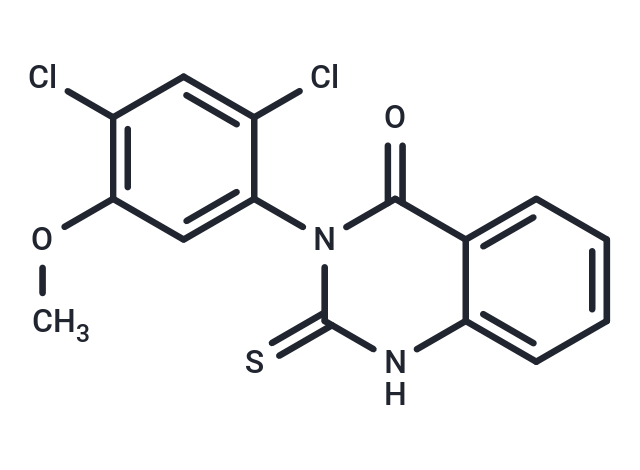
Mdivi-1 (Mitochondrial division inhibitor 1) is a mitochondrial division inhibitor that inhibits DRP1 and Dynamin I (IC50=1-10 μM). Mdivi-1 inhibits mitochondrial autophagy.

| Pack Size | Price | Availability | Quantity |
|---|---|---|---|
| 5 mg | $39 | In Stock | |
| 10 mg | $51 | In Stock | |
| 25 mg | $92 | In Stock | |
| 50 mg | $155 | In Stock | |
| 100 mg | $239 | In Stock | |
| 200 mg | $355 | In Stock | |
| 500 mg | $588 | In Stock | |
| 1 mL x 10 mM (in DMSO) | $43 | In Stock |
| Description | Mdivi-1 (Mitochondrial division inhibitor 1) is a mitochondrial division inhibitor that inhibits DRP1 and Dynamin I (IC50=1-10 μM). Mdivi-1 inhibits mitochondrial autophagy. |
| Targets&IC50 | Dnm1 GTPase:1 μM-10 μM |
| In vitro | In the ganglion cell layer (GCL), Mdivi-1 exhibits strong immunoreactivity. Twelve hours following ischemic induction, Mdivi-1 significantly increases protein expression within the GCL. It notably reduces the expression of GFAP protein without altering Drp1 protein expression. In the normal murine retina, Mdivi-1 primarily localizes to the inner plexiform layer, ganglion cell layer, outer plexiform layer, and inner nuclear layer. Early in the development of the ischemic murine retina, there is a marked increase in the protein expression of Mdivi-1 and glial fibrillary acidic protein (GFAP). Mdivi-1 inhibits apoptosis in the ischemic retina and significantly increases the survival rate of retinal ganglion cells (RGC) two weeks post-ischemia. |
| In vivo | Mdivi-1 inhibits the ATPase activity and self-assembly of Dnm1 by inducing a conformational change (IC50<10 μM). It effectively suppresses C8-Bid and STS-induced mitochondrial outer membrane permeabilization (MOMP) in HeLa cells and extracellular mouse liver mitochondria. Mdivi-1 blocks the divisionof Dynamin-related GTPases, yeast Dnm1, and human Drp1, facilitating efficient and reversible mitochondrial fusion into a net-like structure. Intracellularly, it prevents apoptosis by inhibiting mitochondrial outer membrane permeabilization. Mdivi-1 represents a potential therapeutic class for treating stroke, myocardial infarction, and neurodegenerative diseases. |
| Kinase Assay | All GTPase assay reactions are started in a 200 μL volume, of which 150 μL is placed into the well of a 96-well plate. Depletion of NADH, as monitored by reading the A340 of the reaction, is measured every 20 s for a total of 40 min using a SpectraMAX 250 96-well plate reader. Spectrophotometric data are transferred to Excel and the measured steady state depletion of NADH over time is converted to protein activity. |
| Cell Research | Mdivi-1 is dissolved in DMSO. YPGlycerol plates are topped with 10 mL YPGlycerol containing 1% low melt agar and 75 μM mdivi-1, and cells are spotted 12 hours later using a 48 well pinning device. After pinning cells, plates are incubated at 24°C or 37°C and imaged using an Eagle Eye II imaging system. |
| Alias | Mitochondrial division inhibitor 1 |
| Molecular Weight | 353.22 |
| Formula | C15H10Cl2N2O2S |
| Cas No. | 338967-87-6 |
| Smiles | O=C1N(C(=S)NC=2C1=CC=CC2)C3=CC(OC)=C(Cl)C=C3Cl |
| Relative Density. | 1.57 g/cm3 (Predicted) |
| Storage | store at low temperature | Powder: -20°C for 3 years | In solvent: -80°C for 1 year | Shipping with blue ice. | |||||||||||||||||||||||||
| Solubility Information | DMSO: 50 mg/mL (141.55 mM), Sonication is recommended. 10% DMSO+40% PEG300+5% Tween 80+45% Saline: 3.53 mg/mL (9.99 mM), suspension.In vivo: Please add the solvents sequentially, clarifying the solution as much as possible before adding the next one. Dissolve by heating and/or sonication if necessary. Working solution is recommended to be prepared and used immediately. | |||||||||||||||||||||||||
Solution Preparation Table | ||||||||||||||||||||||||||
DMSO
| ||||||||||||||||||||||||||

Copyright © 2015-2025 TargetMol Chemicals Inc. All Rights Reserved.