- Remove All
 Your shopping cart is currently empty


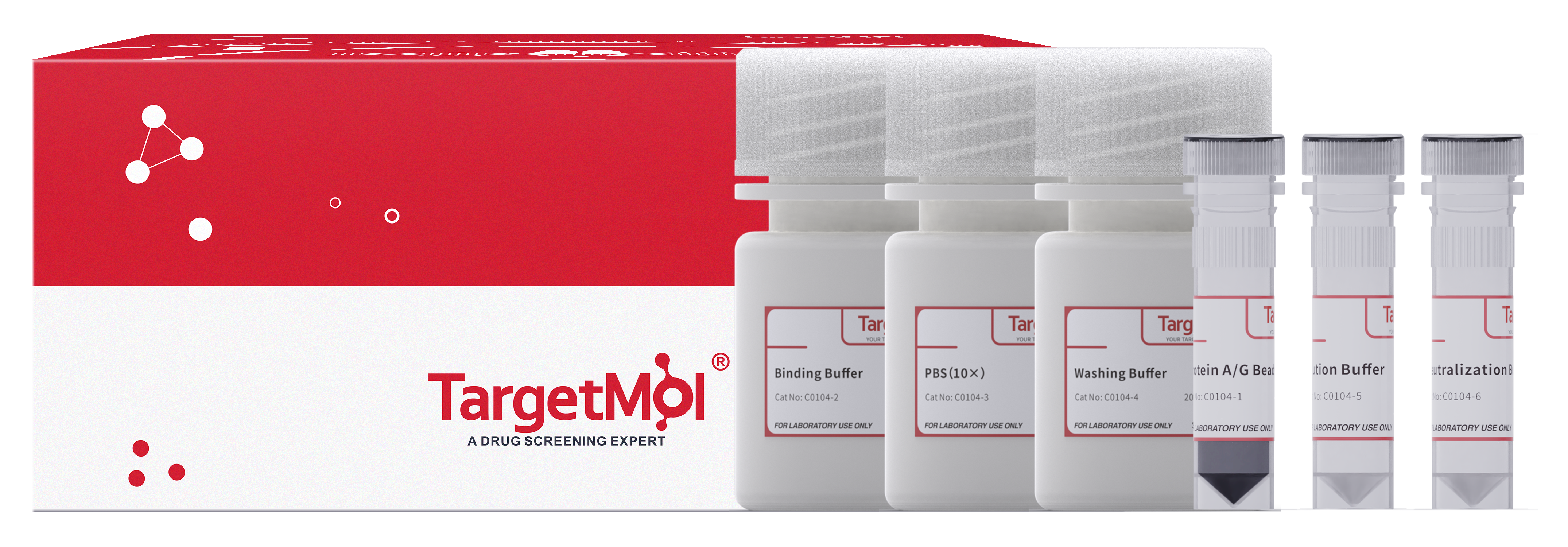
Protein A/G Immunoprecipitation Kit
| Pack Size | Price | Availability | Quantity |
|---|---|---|---|
| 20 T | $140 | In Stock |
 General Information
| Catalog No. | Product Name | Packaging |
|---|---|---|
| C0104-1 | Protein A/G Immunomagnetic Beads | 1 mL |
| C0104-2 | Binding Buffer | 30 mL |
| C0104-3 | PBS(10×) | 20 mL |
| C0104-4 | Washing Buffer | 20 mL |
| C0104-5 | Elution Buffer | 0.5 mL |
| C0104-6 | Neutralization Buffer | 0.2 mL |
| Protein A/G Immunomagnetic Beads | Features |
|---|---|
| Particle Size | 2 μm |
| Human IgG Capacity(Antibody Capacity) | 0.5~0.6 mg/mL |
| Bead Concentration | 10 mg/mL |
| Storage Solution | 1×PBS (0.1%(w/v)BSA+0.1%(V/V)proclin-300) |
| Applicable antibody species | Broad-spectrum antibody species |
| Application | IP , Co-IP , ChIP |
 Product Features
1.Low Non-specific Binding: This product offers more antibody binding sites and demonstrates a low non-specific binding rate.
2.High Efficiency: Each milliliter of Immunomagnetic Beads can bind more than 300 μg of human IgG, and a single immunoprecipitation reaction requires only 25 μL of beads to complete the assay.
3.Simple Operation: The micron-sized beads provide an ultra-large specific surface area, significantly reducing the equilibrium time required for antibody and antigen adsorption. Antibody binding can be completed within 15 minutes, and antigen precipitation can be accomplished within 30 minutes.
4.Reliable Results: Simple operation minimizes the risk of target protein hydrolysis caused by prolonged handling, ensuring the activity of the target protein and the integrity of protein complexes.
 Instruction Mannual
1. Preparation of Antigen Samples
It is recommended to select appropriate pretreatment methods based on the source of the antigen sample to effectively release the target antigen into the sample solution.
(1) Serum Samples: If the target protein is abundant, dilute the serum sample with Binding Buffer to achieve a final target protein concentration of 10–100 µg/mL. Keep the diluted sample on ice for immediate use or store at -20°C for long-term storage.
(2) Under sterile conditions, dilute PBS (10×) to 1×. Centrifuge the cells (4℃, 500 g, 10 min), discard the supernatant, and weigh the pellet. Wash the cells twice with 1× PBS at a ratio of 50 µL per milligram of cells. Then, add Binding Buffer at a ratio of 5–10 µL per milligram of cells, along with a protease inhibitor (1 mM PMSF or Protease Inhibitor Cocktail C0001). Mix well and incubate on ice for 10 minutes. Centrifuge the mixture to collect the supernatant (4℃, 14000 g, 10 min). Keep the supernatant on ice for immediate use or store at -20℃ for long-term preservation.
(3) Adherent Cell Samples: Remove the culture medium and wash the cells twice with 1× PBS at a ratio of 150 µL per 1.0 × 10^5 cells. Use a cell scraper to collect the cells into a 1.5 mL microcentrifuge tube. Add Binding Buffer at a ratio of 20–30 µL per 1.0 × 10^5 cells. Include a protease inhibitor. Mix thoroughly and incubate on ice for 10 minutes. Centrifuge (4°C, 14,000 g, 10 min) and collect the supernatant. Keep on ice for immediate use or store at -20°C for long-term storage.
(4) E. coli Samples: Centrifuge the bacterial culture (4°C, 12,000 g, 2 min) and discard the supernatant. Weigh the bacterial pellet, then wash twice with 1× PBS at a ratio of 10 mL per gram (wet weight) of bacteria. Add Binding Buffer at a ratio of 5–10 mL per gram (wet weight) of bacteria. Include a protease inhibitor. Resuspend the bacteria, then lyse the cells via ultrasonication. Centrifuge (4°C, 17,000 g, 10 min) and collect the supernatant. Keep on ice for immediate use or store at -20°C for long-term storage.
2. Magnetic Bead Pre-treatment
(1) Resuspend the immunomagnetic beads by vortexing for 1 minute. Transfer 25–50 µL of the suspension into a 1.5 mL EP tube.
(2) Add 200 µL of Binding Buffer and place the EP tube into a magnetic stand for 1 minute to perform magnetic separation. Remove the supernatant and EP tube. Repeat this step once.
(3) Resuspend the beads in 200 µL of Binding Buffer for further use.
3. Binding of Antibody
(1) Preparation of Antibody Working Solution: Dilute the antibody sample with Binding Buffer to a final concentration of 5–50 µg/mL. Keep on ice for use.
(2) Antibody Binding: Perform magnetic separation on bead suspension (pre-treated in step 2 and remove the supernatant. Add 200 µL of antibody working solution and resuspend the magnetic beads quickly. Rotate tube or gently pipette at room temperature for 15 minutes. After mixing, perform magnetic separation and collect the supernatant. Keep on ice for further analysis.
(3) Elution: Add 200 µL of Binding Buffer to the tube and gently pipette to disperse the bead-antibody complex. Perform magnetic separation and remove the supernatant. Repeat this step once.
4. Antibody Cross-linking (Optional)
If it is necessary to elute both antibody and target antigen complex, skip this step and proceed directly to Step 5. This step is intended for experiments that require the elution of the target antigen alone. It is recommended to use BS3 as a cross-linker.
5. Immunoprecipitation of Target Antigen
(1) Antigen Binding: Add 200 µL of the antigen sample prepared in Step 1. Gently pipette and to evenly disperse the antigen and magnetic bead-antibody complex. Mix gently at room temperature using a tube rotator or manually invert the EP tube for 10 minutes to ensure efficient antigen-antibody binding. For weaker interactions, incubate at room temperature for 1 hour or at 4°C overnight.
(2) Washing and Transfer: Perform magnetic separation to isolate the magnetic bead-antibody-antigen complex and collect the supernatant, placing it on ice for subsequent analysis. Add 200 µL of Washing Buffer to the EP tube and gently pipette up and down to disperse the magnetic bead-antibody-antigen complex. Perform magnetic separation, discard the supernatant, and remove the EP tube from the magnetic stand. Repeat the washing process twice. Finally, add 200 µL of Washing Buffer, transfer the magnetic bead-antibody-antigen suspension to a new 1.5 mL EP tube, and perform magnetic separation again. Discard the supernatant and remove the EP tube from the magnetic stand.
Note: Before antigen elution, transfer the magnetic beads to a new EP tube to avoid co-eluting nonspecifically adsorbed proteins from the tube walls.
6. Elution
Two elution methods are recommended.
(1) Denaturation Elution Method: suitable for SDS-PAGE analysis. Add 25 µL of 1× SDS-PAGE Loading Buffer to the EP tube, mix thoroughly, and heat at 95°C for 5 minutes. Perform magnetic separation or centrifugation (at room temperature, 13000 g, 10 minutes). Collect the supernatant, and proceed with SDS-PAGE analysis.
(2) Non-Denaturation Elution Method: This method preserves the biological activity of the eluted samples. Add 20 µL of Elution Buffer to the EP tube, mix thoroughly, and incubate at room temperature for 10 minutes. Perform magnetic separation or centrifugation (room temperature, 13000 g, 10 minutes). Transfer the supernatant to a new EP tube, and immediately add 1.0 µL of Neutralization Buffer to adjust the pH of the eluted product to neutral.
 Storage
Store at 4℃ for 2 years.
 Precaution
1.Do not dry or freeze the magnetic beads.
2.To minimize bead loss, magnetic separation is recommended to last more than 1 minute.
3.Shake fully before removing the beads from the tube to ensure uniform suspension. Bubbles should be avoided during operation.
4.It is recommended to use high-quality pipette tips and reaction tubes to minimize loss due to bead or solution adhesion.
5.PBS (10×) should be diluted under sterile conditions. If contamination of the solution is detected, discontinue use immediately.
6.For optimal experimental results, select antibodies with high specificity for immunoprecipitation (IP) reactions.
7.Based on experimental needs, the operator may utilize the supernatants collected during the antibody binding and antigen binding steps to evaluate the interactions between antibodies, antigens, and magnetic beads.
8.In IP experiments, the affinity between different types of antibodies and antigens may vary. The composition of the Binding Buffer and Washing Buffer also influences antibody-antigen interactions. If the recommended buffer system does not yield satisfactory results, operators can optimize and prepare custom buffer systems.
9.Although the recombinant Protein A/G coating on the bead surface remains stable under extreme conditions (e.g., low pH, heat treatment), it is not recommended for immunoprecipitation experiments targeting proteins with molecular weights around 130 kDa.
10.This product is for R&D use only, not for diagnostic procedures, food, drug, household, or other uses.
11.It’s advisable to wear a lab coat and disposable glove.
 Instruction Manual

Copyright © 2015-2025 TargetMol Chemicals Inc. All Rights Reserved.


