Shopping Cart
- Remove All
 Your shopping cart is currently empty

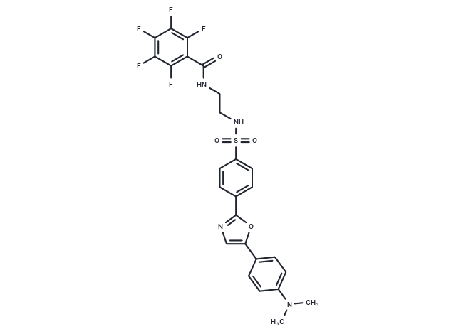
ER-Tracker dye, a derivative of BODIPY series dyes coupled with Glibenclamide, exhibits high selectivity for the endoplasmic reticulum and is non-toxic to cells at low concentrations. This environmentally sensitive probe retains part of its fluorescence after formaldehyde treatment and features high fluorescence life and a good extinction coefficient. Glibenclamide, an ATP-dependent K+ channel blocker (Kir6, KATP) and CFTR Cl-channel blocker, binds in the endoplasmic reticulum. However, ER-Tracker is not suitable for staining cells after fixation [1].

| Pack Size | Price | Availability | Quantity |
|---|---|---|---|
| 10 mg | Inquiry | Inquiry | |
| 50 mg | Inquiry | Inquiry |
| Description | ER-Tracker dye, a derivative of BODIPY series dyes coupled with Glibenclamide, exhibits high selectivity for the endoplasmic reticulum and is non-toxic to cells at low concentrations. This environmentally sensitive probe retains part of its fluorescence after formaldehyde treatment and features high fluorescence life and a good extinction coefficient. Glibenclamide, an ATP-dependent K+ channel blocker (Kir6, KATP) and CFTR Cl-channel blocker, binds in the endoplasmic reticulum. However, ER-Tracker is not suitable for staining cells after fixation [1]. |
| In vitro | ER-Tracker solution preparation involves first creating a storage solution by diluting 100 μg of ER-Tracker with 172 μL of anhydrous DMSO to obtain a 1 mM stock solution. The storage solution should be aliquoted and stored at -20℃ or -80℃, protected from light. Next, prepare the working solution by diluting the stock solution with pre-warmed serum-free cell culture medium or PBS to concentrations between 100 nM and 1 μM, adjusting the concentration based on specific requirements, and using it immediately after preparation. For staining suspension cells, centrifuge to collect the cells and wash twice with PBS, five minutes each time, maintaining a cell density of 1×10^6/mL. Add 1 mL of ER-Tracker working solution and incubate at room temperature for 5-30 minutes. Centrifuge at 400 g for 3-4 minutes, discard the supernatant, and wash the cells twice with PBS for five minutes each. Resuspend cells in 1 mL serum-free medium or PBS, then observe using a fluorescence microscope or flow cytometer. For staining adherent cells, culture on sterile coverslips, remove coverslips from the medium, and aspirate excess medium. Add 100 μL of dye working solution, gently agitate to cover cells completely, and incubate for 5-30 minutes. Aspirate the working solution and wash 2-3 times with medium for five minutes each, observing with a fluorescence microscope. For flow cytometry analysis, cells need to be detached with trypsin, resuspended, and then stained. |
| Molecular Weight | 580.53 |
| Formula | C26H21F5N4O4S |
| Cas No. | 287715-95-1 |
| Storage | Powder: -20°C for 3 years | In solvent: -80°C for 1 year | Shipping with blue ice. |

Copyright © 2015-2025 TargetMol Chemicals Inc. All Rights Reserved.