Shopping Cart
- Remove All
 Your shopping cart is currently empty

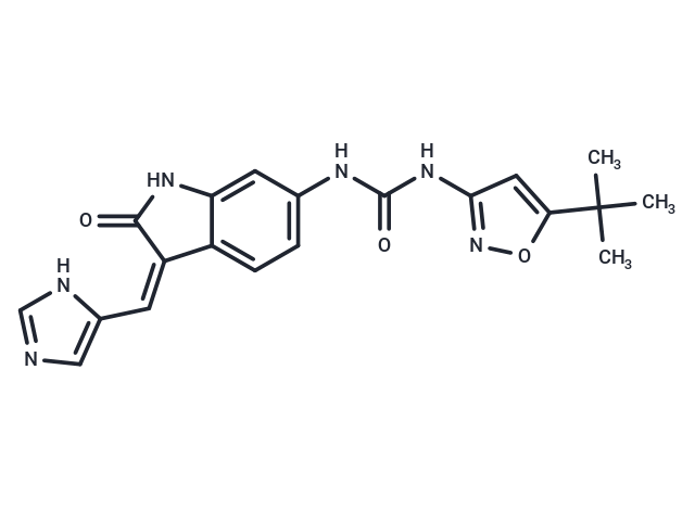
CSF1R-IN-22 (Compound C19), a potent orally administered CSF-1R selective inhibitor (IC50 <6 nM), significantly enhances CXCL9 secretion from M2 macrophages and promotes CD8+ T cell infiltration. Moreover, it amplifies the anti-tumor immune responses in conjunction with anti-PD-1 and triggers apoptosis in tumor cells. Additionally, CSF1R-IN-22 effectively reprograms M2-like TAMs (tumor-associated macrophages) to an M1 phenotype, modulates the tumor microenvironment (TME) by fostering the recruitment of CD8+ T cells, and diminishes the presence of immunosuppressive Tregs and MDSCs [1].

| Pack Size | Price | Availability | Quantity |
|---|---|---|---|
| 10 mg | Inquiry | 10-14 weeks | |
| 50 mg | Inquiry | 10-14 weeks |
| Description | CSF1R-IN-22 (Compound C19), a potent orally administered CSF-1R selective inhibitor (IC50 <6 nM), significantly enhances CXCL9 secretion from M2 macrophages and promotes CD8+ T cell infiltration. Moreover, it amplifies the anti-tumor immune responses in conjunction with anti-PD-1 and triggers apoptosis in tumor cells. Additionally, CSF1R-IN-22 effectively reprograms M2-like TAMs (tumor-associated macrophages) to an M1 phenotype, modulates the tumor microenvironment (TME) by fostering the recruitment of CD8+ T cells, and diminishes the presence of immunosuppressive Tregs and MDSCs [1]. |
| In vitro | CSF1R-IN-22 significantly inhibits the activation of the CSF-1R signaling pathway in BMDMs cells at concentrations of 0-2500 nM for 1 hour. At concentrations of 30-100 nM for 24 hours, CSF1R-IN-22 effectively reprograms M2 macrophages into M1 macrophages in both BMDMs and HMDMs cells. The supernatant from M2 macrophages treated with 10-100 nM of CSF1R-IN-22 for 20 hours significantly reduces the viability of MC-38 and CT-26 cells. A Western blot analysis using BMDMs cells at concentrations of 10, 30, and 100 nM for 1 hour showed a dose-dependent inhibition of the phosphorylation of CSF-1R and its downstream signaling proteins AKT and mTORC1. An apoptosis analysis with MC-38 and CT-26 cells at 10, 30, and 100 nM for 20 hours demonstrated that the processed M2 macrophage supernatant substantially increased the apoptosis rate, with an approximately 60% increase observed at the 100 nM concentration. |
| In vivo | CSF1R-IN-22 exhibited significant antitumor activity in C57BL/6 mice bearing MC-38 subcutaneous tumors at dosages of 5-20 mg/kg, orally once daily for 14 days. It enhanced the activity of cytotoxic T lymphocytes (CTLs), with high-dose groups outperforming PLX3397 [1]. When administered in combination with 100 μg/mouse PD-1 antibody, CSF1R-IN-22 (20 mg/kg; p.o.; once daily for 14 days) demonstrated enhanced antitumor effects. A positive correlation was observed between CXCL9 expression and survival rates [1]. Pharmacokinetic analysis in SD rats [1] showed that intravenous administration of 1 mg/kg resulted in an AUC 0-t of 9126.62 ng·h/mL, a half-life of 1.34 hours, a clearance rate of 0.10 L/h/kg, and a steady-state volume of distribution of 0.34 L/kg; oral administration of 10 mg/kg resulted in an AUC 0-t of 85939.36 ng·h/mL, a half-life of 2.41 hours, a clearance rate of 0.12 L/h/kg, and a maximum concentration of 10867.65 ng/mL, with a bioavailability of 94.2%. In the MC-38 tumor model in C57BL/6 mice, CSF1R-IN-22 significantly inhibited tumor growth and induced apoptosis through oral administration of 5, 10, 20 mg/kg daily for 14 days, leading to significantly lower tumor mass compared to controls. The high-dose group showed a tumor growth inhibition (TGI) of 65% and a 30-day survival rate of 70%. An increase in M1 macrophage marker gene mRNA levels (Nos2, Tnf, Il6, Il1) and a decrease in M2 macrophage marker gene expression (Arg1, Chil3l, Rentla, Mrc1) were observed. Doses of 10 and 20 mg/kg significantly increased the proportion of CD3+CD8+ T cells, and reduced the proportion of immunosuppressive Treg cells and myeloid-derived suppressor cells (MDSCs) in tumor tissues. For mice treated with CSF1R-IN-22 (20 mg/kg) in combination with PD-1 in the MC-38 or MC-38-luc models, there was a significant reduction in tumor bioluminescence compared to the monotherapy groups. The combination therapy notably increased the proportion of CD3+CD8+ T cells and CTLs in tumor tissues, significantly decreasing the proportion of immunosuppressive Treg cells in the MC-38 tumor model. Enhanced infiltration of CD8+ T cells and CTLs was significantly associated with increased mRNA expression of Cxcl9. Mice treated with the combination therapy exhibited a survival rate of 100% at 70 days and 70% overcame tumor recurrence within 91 days after rechallenging MC-38 tumor cells. The use of a CXCL9 neutralizing antibody significantly reduced apoptosis and the proportion of CD3+CD8+ T cells. |
| Molecular Weight | 392.41 |
| Formula | C20H20N6O3 |
| Cas No. | 2760585-35-9 |
| Storage | Powder: -20°C for 3 years | In solvent: -80°C for 1 year | Shipping with blue ice. |

Copyright © 2015-2025 TargetMol Chemicals Inc. All Rights Reserved.