- Remove All
 Your shopping cart is currently empty
AMH Protein, Human, Recombinant (His)
Anti-Mullerian hormone (AMH), a member of the TGF-beta superfamily, is produced by granulosa cells (GCs) of preantral and small antral follicles and plays a role in regulating the recruitment of primordial follicles and the FSH-dependent development of follicles. BMP15 up-regulates the transcription of AMH and that the inhibition of p38 MAPK decreases the BMP15-induced expression of AMH and SOX9, suggesting that BMP15 up-regulates the expression of AMH via the p38 MAPK signaling pathway, and this process involves the SOX9 transcription factor. AMH is widely used for assessing ovarian reserve, and it is particularly convenient, because it is thought to have minimal variability throughout the menstrual cycle. Fetal anti-Mullerian hormone (AMH) is responsible for normal male sexual differentiation, and circulating AMH is used as a marker of testicular tissue in newborns with disorders of sex development. Anti-Mullerian hormone (AMH) produced in the developing testis induces the regression of the Mullerian duct, which develops into the oviducts, uterus and upper vagina. As well as other hormone receptors, and a decreased ovarian cortex cell proliferation. These results help understand the inhibitory effects of AMH on follicular development.

AMH Protein, Human, Recombinant (His)
| Pack Size | Price | Availability | Quantity |
|---|---|---|---|
| 50 μg | $386 | In Stock | |
| 100 μg | $660 | 7-10 days | |
| 200 μg | $1,120 | 7-10 days | |
| 500 μg | $2,270 | 7-10 days |
Product Information
| Biological Activity | Activity testing is in progress. It is theoretically active, but we cannot guarantee it. If you require protein activity, we recommend choosing the eukaryotic expression version first. |
| Description | Anti-Mullerian hormone (AMH), a member of the TGF-beta superfamily, is produced by granulosa cells (GCs) of preantral and small antral follicles and plays a role in regulating the recruitment of primordial follicles and the FSH-dependent development of follicles. BMP15 up-regulates the transcription of AMH and that the inhibition of p38 MAPK decreases the BMP15-induced expression of AMH and SOX9, suggesting that BMP15 up-regulates the expression of AMH via the p38 MAPK signaling pathway, and this process involves the SOX9 transcription factor. AMH is widely used for assessing ovarian reserve, and it is particularly convenient, because it is thought to have minimal variability throughout the menstrual cycle. Fetal anti-Mullerian hormone (AMH) is responsible for normal male sexual differentiation, and circulating AMH is used as a marker of testicular tissue in newborns with disorders of sex development. Anti-Mullerian hormone (AMH) produced in the developing testis induces the regression of the Mullerian duct, which develops into the oviducts, uterus and upper vagina. As well as other hormone receptors, and a decreased ovarian cortex cell proliferation. These results help understand the inhibitory effects of AMH on follicular development. |
| Species | Human |
| Expression System | HEK293 Cells |
| Tag | N-His |
| Accession Number | P03971 |
| Synonyms | MIS,MIF,anti-Mullerian hormone |
| Construction | A DNA sequence encoding the human AMH (NP_000470.2) (Leu25-Arg560) was expressed with a polyhistidine tag at the N-terminus. Predicted N terminal: His |
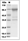
| Protein Purity | > 90 % as determined by SDS-PAGE.  |
| Molecular Weight | 59.1 kDa (predicted); 63.6, 40.5, 35 kDa (reducing conditions) |
| Endotoxin | < 1.0 EU/μg of the protein as determined by the LAL method. |
| Formulation | Lyophilized from a solution filtered through a 0.22 μm filter, containing 20 mM Tris, 150 mM NaCl, pH 8.0.Typically, a mixture containing 5% to 8% trehalose, mannitol, and 0.01% Tween 80 is incorporated as a protective agent before lyophilization. |
| Reconstitution | A Certificate of Analysis (CoA) containing reconstitution instructions is included with the products. Please refer to the CoA for detailed information. |
| Stability & Storage | It is recommended to store recombinant proteins at -20°C to -80°C for future use. Lyophilized powders can be stably stored for over 12 months, while liquid products can be stored for 6-12 months at -80°C. For reconstituted protein solutions, the solution can be stored at -20°C to -80°C for at least 3 months. Please avoid multiple freeze-thaw cycles and store products in aliquots. |
| Shipping | In general, Lyophilized powders are shipping with blue ice. |
| Research Background | Anti-Mullerian hormone (AMH), a member of the TGF-beta superfamily, is produced by granulosa cells (GCs) of preantral and small antral follicles and plays a role in regulating the recruitment of primordial follicles and the FSH-dependent development of follicles. BMP15 up-regulates the transcription of AMH and that the inhibition of p38 MAPK decreases the BMP15-induced expression of AMH and SOX9, suggesting that BMP15 up-regulates the expression of AMH via the p38 MAPK signaling pathway, and this process involves the SOX9 transcription factor. AMH is widely used for assessing ovarian reserve, and it is particularly convenient, because it is thought to have minimal variability throughout the menstrual cycle. Fetal anti-Mullerian hormone (AMH) is responsible for normal male sexual differentiation, and circulating AMH is used as a marker of testicular tissue in newborns with disorders of sex development. Anti-Mullerian hormone (AMH) produced in the developing testis induces the regression of the Mullerian duct, which develops into the oviducts, uterus and upper vagina. As well as other hormone receptors, and a decreased ovarian cortex cell proliferation. These results help understand the inhibitory effects of AMH on follicular development. |
Dose Conversion
Calculator
Tech Support

Copyright © 2015-2025 TargetMol Chemicals Inc. All Rights Reserved.


