Shopping Cart
- Remove All
 Your shopping cart is currently empty

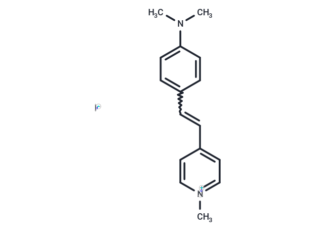
4-Di-1-ASP, a styryl dye, selectively stains glioma cells within live brain tissue, facilitating microscopic analysis of cell structure, viability, and processes including proliferation, endocytosis, cytokinesis, and phagocytosis. It also allows for the observation of mitochondrial structures in living cells. The dye exhibits green fluorescence when visualized under a microscope (λ ex /λ em = 475/606 nm) [1] [2].


| Description | 4-Di-1-ASP, a styryl dye, selectively stains glioma cells within live brain tissue, facilitating microscopic analysis of cell structure, viability, and processes including proliferation, endocytosis, cytokinesis, and phagocytosis. It also allows for the observation of mitochondrial structures in living cells. The dye exhibits green fluorescence when visualized under a microscope (λ ex /λ em = 475/606 nm) [1] [2]. |
| In vitro | Guidelines (Following is our recommended protocol. This protocol only provides a guideline, and should be modified according to your specific needs) [1]. Prepare a 2 mM stock solution of the dye in water. Incubate cell sections at 35°C and add the dye to achieve a final concentration of 1 μM, staining for 10 minutes. After staining, mount the sections on a microscope slide, cover with a coverslip, and examine under a fluorescent confocal microscope with peak excitation and emission wavelengths at 475 nm and 606 nm, respectively. |
| Molecular Weight | 366.24 |
| Formula | C16H19IN2 |
| Cas No. | 959-81-9 |
| Storage | keep away from direct sunlight | Shipping with blue ice. |

Copyright © 2015-2025 TargetMol Chemicals Inc. All Rights Reserved.