
Shopping Cart
- Remove All
 Your shopping cart is currently empty

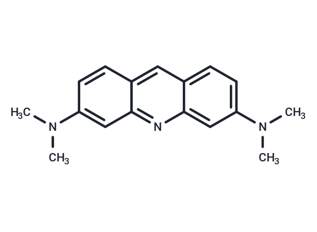
Acridine Orange base is a cell-permeable fluorescent dye that binds to nucleic acids and emits different colors of fluorescence, which exhibits green fluorescence when bound to dsDNA (Ex=488 nm, Em=520-524 nm) and red fluorescence when bound to ssDNA or ssRNA (Ex=457 nm, Em=630-644 nm) and can be used for cell cycle analysis and apoptosis detection.
| Pack Size | Price | Availability | Quantity |
|---|---|---|---|
| 50 mg | $30 | In Stock | |
| 100 mg | $42 | In Stock | |
| 500 mg | $98 | In Stock | |
| 1 g | $143 | In Stock |
| Description | Acridine Orange base is a cell-permeable fluorescent dye that binds to nucleic acids and emits different colors of fluorescence, which exhibits green fluorescence when bound to dsDNA (Ex=488 nm, Em=520-524 nm) and red fluorescence when bound to ssDNA or ssRNA (Ex=457 nm, Em=630-644 nm) and can be used for cell cycle analysis and apoptosis detection. |
| In vitro | Fixed cell staining I. Sample preparation: 1. For cells in suspension culture or hematology samples: rinse cells once with ice-cold PBS and suspend in ice-cold PBS at 10^6 cells/mL. 2. For cells attached to tissue culture plates: collect cells from flasks or culture dishes by trypsinization, fill the trypsinized cells with cells floating in the culture medium (mainly isolated mitotic and dead cells), and then use culture medium containing serum to inactivate trypsin. Finally, suspend the cells in ice-cold PBS at about 10^6 cells/mL. 3. For cells isolated from solid tumors: rinse cells without any enzymes for cell dissociation and suspend in ice-cold PBS at about 10^6 cells/mL. II. Operation steps: 1. Transfer 1 mL of cell suspension with a Pasteur pipette to a 15 mL conical glass tube containing 10 mL of ice-cold 70% ethanol. Fix the cells on ice for ≥2 hours. 2. Centrifuge the tube at 300 × g, 4°C for 5 min. Remove all ethanol with ice-cold PBS, rinse once, and suspend in ice-cold PBS at a density of <2×10^6 cells/mL. 3. Take 0.2 mL of cell suspension (≤2×10^5 cells) and transfer to a small tube. Cool on ice. 4. Add 0.4 mL of ice-cold permeabilization solution. Wait 15 seconds and keep cells on ice. 5. Add 1.2 mL of ice-cold AO staining solution. Keep cells on ice. 6. Add 1.2 mL of ice-cold Acridine Orange base staining solution. Keep cells on ice. 7.After adding Acridine Orange base solution, measure and record cell fluorescence using a flow cytometer within 2 to 10 minutes. [1] |
| Molecular Weight | 265.35 |
| Formula | C17H19N3 |
| Cas No. | 494-38-2 |
| Smiles | CN(C)c1ccc2cc3ccc(cc3nc2c1)N(C)C |
| Storage | keep away from direct sunlight | store at 4°C | Shipping with blue ice. | ||||||||||||||||||||||||||||||||||||||||
| Solubility Information | DMSO: 30 mg/mL (113.06 mM), Sonication is recommended. H2O: 4 mg/mL (15.07 mM), Sonication is recommended. 1 M HCL: 180 mg/mL (678.35 mM), Sonication is recommended. | ||||||||||||||||||||||||||||||||||||||||
Solution Preparation Table | |||||||||||||||||||||||||||||||||||||||||
H2O/DMSO/1 M HCL
DMSO/1 M HCL
| |||||||||||||||||||||||||||||||||||||||||
 For example, your dosage is 10 mg/kg Each animal weighs 20 g, and the dosage volume is 100 μL . A total of 10 animals were administered, and the formula you used is 5%
  DMSO+30% PEG300+5% Tween 80+60% ddH2O. So your working solution concentration is 2 mg/mL。 (mother liquor concentration of 40 mg/mL), if you need to configure a concentration that exceeds the solubility of the product, please contact us first. main solution, add 300 μLPEG300 mix well and clarify, then add 50 more μL Tween 80, mix well and clarify, then add 600 more μLddH2O mix well and clarify
 Hello! How can I help you today?
Copyright © 2015-2025 TargetMol Chemicals Inc. All Rights Reserved.