
Shopping Cart
- Remove All
 Your shopping cart is currently empty

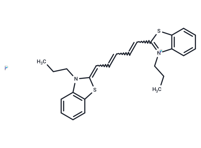
DiSC3(5) is a lipophilic cationic fluorescent probe sensitive to changes in membrane potential. 3,3'-Dipropylthiadicarbocyanine iodide accumulates in hyperpolarized cell membranes, leading to fluorescence quenching; when the membranes are depolarized, the dye is released from the cells and the fluorescence intensity is enhanced, which is used for real-time monitoring of mitochondrial membrane potential changes and membrane hyperpolarization. The maximum excitation/emission wavelength was 622/670 nm.
| Pack Size | Price | Availability | Quantity |
|---|---|---|---|
| 25 mg | $42 | In Stock | |
| 50 mg | $68 | In Stock | |
| 100 mg | $118 | In Stock | |
| 500 mg | $298 | In Stock |
| Description | DiSC3(5) is a lipophilic cationic fluorescent probe sensitive to changes in membrane potential. 3,3'-Dipropylthiadicarbocyanine iodide accumulates in hyperpolarized cell membranes, leading to fluorescence quenching; when the membranes are depolarized, the dye is released from the cells and the fluorescence intensity is enhanced, which is used for real-time monitoring of mitochondrial membrane potential changes and membrane hyperpolarization. The maximum excitation/emission wavelength was 622/670 nm. |
| In vitro | Instructions I. Solution preparation 1. Stock solution preparation: Stock solutions are prepared at 1-5 mM in DMSO or EtOH. Note: The unused portion of the stock solution should be stored at -20°C. Avoid repeated freezing and thawing. 2. Working solution preparation: Dilute the stock solution into a suitable buffer, such as pure DMEM, HBSS or PBS, to make a 1 to 5 uM working solution. Note: The concentration of the working solution should be determined empirically for different cell types and/or experimental conditions. It is recommended to test at concentrations over a range of at least ten times. II. Operation steps 1. Staining suspended cells: 1) Suspend cells in the dye working solution at a density of 1×10^6/mL. 2) Incubate at 37°C for 2-20 minutes. The incubation time depends on the cell type. Incubate for 20 minutes first, then optimize as needed to obtain uniform labeling. 3) Centrifuge the labeled suspension tube at 1000 to 1500 rpm for 5 minutes. 4) Remove the supernatant and gently resuspend the cells in pre-warmed (37°C) growth medium. 5) Wash twice. 2. Staining adherent cells: 1) Grow adherent cells on sterile glass coverslips. 2) Remove the coverslips from the growth medium and gently drain the excess medium. Place the coverslips in a humidity chamber. 3) Pipette 100 μL of the dye working solution into the corner of the coverslip and gently agitate until all cells are covered. 4) Incubate the coverslips at 37°C for 2-20 minutes. Incubation time varies depending on cell type. Start with a 20-minute incubation and optimize as needed to obtain uniform labeling. 5) Drain the dye working solution and wash the coverslips two to three times with growth medium. For each wash cycle, cover the cells with pre-warmed growth medium, incubate for 5-10 minutes, and then drain the medium. III. Microscope detection. IV. Flow cytometer detection: Cells labeled with DiS can be analyzed using the conventional FL3 flow cytometer detection channel. |
| Alias | 3,3'-Dipropylthiadicarbocyanine iodide |
| Molecular Weight | 546.53 |
| Formula | C25H27IN2S2 |
| Cas No. | 53213-94-8 |
| Smiles | [I-].S1C=2C=CC=CC2N(C1=CC=CC=CC=3SC=4C=CC=CC4[N+]3CCC)CCC |
| Storage | keep away from direct sunlight | Powder: -20°C for 3 years | In solvent: -80°C for 1 year | Shipping with blue ice. | |||||||||||||||
| Solubility Information | DMSO: 4 mg/mL (7.32 mM), Sonication is recommended. | |||||||||||||||
Solution Preparation Table | ||||||||||||||||
DMSO
| ||||||||||||||||
 For example, your dosage is 10 mg/kg Each animal weighs 20 g, and the dosage volume is 100 μL . A total of 10 animals were administered, and the formula you used is 5%
  DMSO+30% PEG300+5% Tween 80+60% ddH2O. So your working solution concentration is 2 mg/mL。 (mother liquor concentration of 40 mg/mL), if you need to configure a concentration that exceeds the solubility of the product, please contact us first. main solution, add 300 μLPEG300 mix well and clarify, then add 50 more μL Tween 80, mix well and clarify, then add 600 more μLddH2O mix well and clarify
 Hello! How can I help you today?
Copyright © 2015-2025 TargetMol Chemicals Inc. All Rights Reserved.