Shopping Cart
- Remove All
 Your shopping cart is currently empty

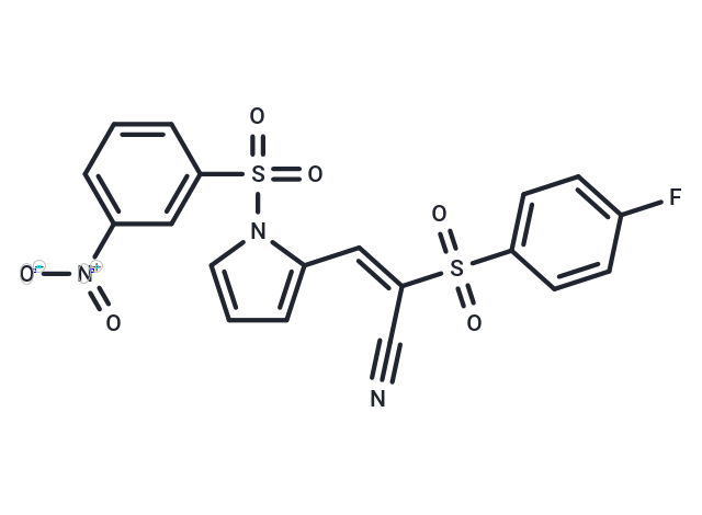
AMZ30 is a selective inhibitor of PME-1 (IC50: 600 nM). It also reduces the demethylated form of PP2A in living cells.

| Pack Size | Price | Availability | Quantity |
|---|---|---|---|
| 1 mg | $32 | In Stock | |
| 5 mg | $70 | In Stock | |
| 10 mg | $108 | In Stock | |
| 25 mg | $183 | In Stock | |
| 50 mg | $277 | In Stock | |
| 100 mg | $410 | In Stock | |
| 1 mL x 10 mM (in DMSO) | $118 | In Stock |
| Description | AMZ30 is a selective inhibitor of PME-1 (IC50: 600 nM). It also reduces the demethylated form of PP2A in living cells. |
| Targets&IC50 | PME-1:600 nM |
| In vitro | Incubation of HEK 293T cells with a concentration range of AMZ30 generated an in situ IC50 value for PME-1 inhibition of 3.5 μM. In HEK 293T cells stably overexpressing PME-1, AMZ30 (20 μM) caused an ~80% reduction in the levels of demethylated PP2A. AMZ30 treatment also significantly increased the methylated form of PP2A. AMZ30 (20 μM) also decreased the demethylated form of PP2A to a similar degree in untransfected HEK 293T cells expressing basal levels of PME-1. |
| Kinase Assay | Purified wild-type PME-1 (500 nM, 2.6 mL total volume in PBS) was incubated with DMSO or AMZ30 (50 μM) at 25 °C. After 30 min, 100 μL was removed from each reaction (Fraction A). The remaining 2.5 mL of each reaction was passaged over a Sephadex G-25M column and eluted in a volume of 3.5 mL PBS (Fraction B). 100 μL of both fractions A and B were labeled with FP-rhodamine (2 μM). After 30 min, the reactions were quenched with 2× SDS-PAGE loading buffer, separated by SDS-PAGE, and analyzed by in-gel fluorescence scanning. Gels were then subjected to Coomassie staining with InstantBlue to verify equivalent protein loading. |
| Cell Research | HEK 293T cells were grown in DMEM SILAC media supplemented with dialyzed fetal bovine serum and 12C14N-lysine and - arginine for "light" cells or 13C615N2-lysine and -arginine for "heavy" cells. Cells were treated as indicated with DMSO or 28 (1 h, 37 °C), washed, harvested, and soluble and membrane proteomes were isolated as described above. Light and heavy proteome fractions (0.5 mg each) were combined (1 mL total volume) and labeled with 5 μM of FP-biotin for 1 hr at 25 °C. After incubation, the membrane proteomes were solubilized with 1% Triton-X and rotated at 4 °C for 1 hr. Enrichment of FP-labeled proteins was achieved as previously described.34 The streptavidin-enriched proteome was washed two times for 3 min with (1) 1% SDS in PBS, (2) 6 M urea in PBS, (3) PBS (pH 7.5) and finally resuspended in 200 μL 8 M urea in 25 mM ammonium bicarbonate. Samples were then prepared for on-bead digestion by reduction with 10 mM TCEP for 30 min at 25 °C and alkylation with 12 mM iodoacetamide for 30 min at 25 °C in the dark. Samples were diluted to 2 M urea with PBS (pH 7.5) and digestions were performed for 12 hr at 37 °C with sequence-grade modified trypsin (4 μL of 0.5 μg/μL) in the presence of 2 mM CaCl2. Lastly, peptide samples were acidified with formic acid to a final concentration of 5%. |
| Molecular Weight | 461.44 |
| Formula | C19H12FN3O6S2 |
| Cas No. | 1313613-09-0 |
| Smiles | [O-][N+](=O)c1cccc(c1)S(=O)(=O)n1cccc1\C=C(/C#N)S(=O)(=O)c1ccc(F)cc1 |
| Relative Density. | no data available |
| Storage | Powder: -20°C for 3 years | In solvent: -80°C for 1 year | Shipping with blue ice. | |||||||||||||||||||||||||
| Solubility Information | DMSO: 22.5 mg/mL (48.76 mM) | |||||||||||||||||||||||||
Solution Preparation Table | ||||||||||||||||||||||||||
DMSO
| ||||||||||||||||||||||||||

Copyright © 2015-2024 TargetMol Chemicals Inc. All Rights Reserved.