Shopping Cart
- Remove All
 Your shopping cart is currently empty

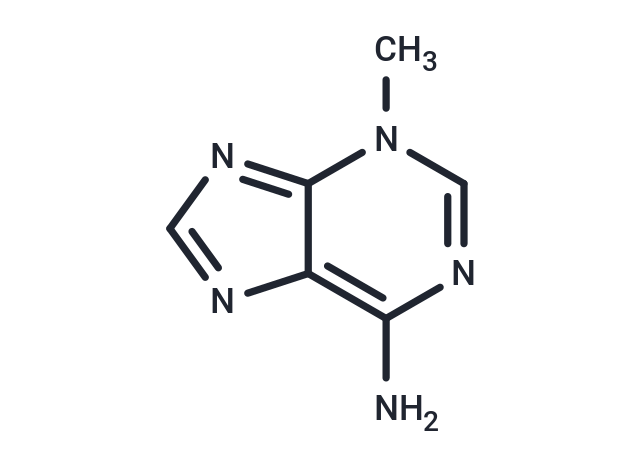
3-Methyladenine (3-MA) is a PI3K inhibitor that selectively inhibits class IB PI3Kγ (IC50 = 60 μM) and class III VPS34 (IC50 = 25 μM). 3-Methyladenine inhibits autophagy.

| Pack Size | Price | Availability | Quantity |
|---|---|---|---|
| 25 mg | $35 | In Stock | |
| 50 mg | $48 | In Stock | |
| 100 mg | $81 | In Stock | |
| 200 mg | $140 | In Stock | |
| 500 mg | $266 | In Stock |
| Description | 3-Methyladenine (3-MA) is a PI3K inhibitor that selectively inhibits class IB PI3Kγ (IC50 = 60 μM) and class III VPS34 (IC50 = 25 μM). 3-Methyladenine inhibits autophagy. |
| Targets&IC50 | PI3Kγ:60 μM (in HeLa cells), VPS34:25 μM (in HeLa cells) |
| In vitro | METHODS: Human cervical cancer cells HeLa were treated with 3-Methyladenine (2.5-10 mM) for 48 h. Cell growth inhibition was detected by Trypan blue dye exclusion assay. RESULTS: 3-Methyladenine decreased HeLa cell viability in a time- and dose-dependent manner. [1] METHODS: Adipocytes 3T3-L1 were treated with 3-Methyladenine (5 mM) for 4 h in the absence of serum, and the expression levels of target proteins were detected by Western Blot. RESULTS: 3-Methyladenine significantly decreased the intracellular level of LC3-II, a marker of autophagy, and increased the expression of p62, indicating that 3-Methyladenine was effective in inhibiting autophagy. [2] METHODS: Mouse melanoma cells B16 were treated with 2DG (5 mM), rotenone (1 μM) and 3-Methyladenine (1.2-5 mM) for 24 h. Cytotoxicity was detected by LDH release assay. RESULTS: 3-Methyladenine dose-dependently reduced the up-regulation of LDH release induced by 2DG/rotenone. 3-Methyladenine protected tumor cells from inhibition of glycolysis and mitochondrial respiration. [3] |
| In vivo | METHODS: To investigate the effects of 3-Methyladenine on atherosclerosis, 3-Methyladenine (30 mg/kg) was injected intraperitoneally into HFD-fed ApoE-/- mice twice weekly for eight weeks. RESULTS: In mice fed a high-fat diet, 3-Methyladenine treatment significantly reduced the size of atherosclerotic plaques and increased the stability of the lesions. 3-Methyladenine has multiple atheroprotective effects on atherosclerosis, including modulation of macrophage autophagy and foam cell formation as well as alteration of the immune microenvironment. [4] METHODS: To investigate the regulatory role of autophagy, a single dose of 3-Methyladenine (15 mg/kg ) was administered intraperitoneally to LPS-induced endotoxic shock in C57/BL6 mice. RESULTS: Animals treated with LPS in combination with 3-Methyladenine showed increased survival and decreased serum inflammatory mediators TNF-α and IL-6 after endotoxemia. [5] |
| Cell Research | Cells were seeded in an 8-well coverglass-bottomed chamber for 24 hours (6×10^3 cells per well). Images were acquired automatically at multiple locations on the coverglass using a Nikon TE2000E inverted microscope fitted with a 20× Nikon Plan Apo objective, a linearly-encoded stage, and a Hamamatsu Orca-ER CCD camera. A mercury-arc lamp with two neutral density filters (for a total 128-fold reduction in intensity) was used for fluorescence illumination. The microscope was controlled using NIS-Elements Advanced Research software and housed in a custom-designed 37°C chamber with a secondary internal chamber that delivered humidified 5% CO2. Fluorescence and differential interference contrast images were obtained every 10 min for a period of 48 hours. To analyze live cell imaging movies, the time-lapse records of live cell imaging experiments were exported as an image series and analyzed manually using NIS-Elements Advanced Research software. The criteria for analyses were described previously, and lagging chromosomes in prometaphase were defined as the red fluorescence-positive materials that lingered outside the roughly formed metaphase plate for more than 3 frames (30 min) [2]. |
| Animal Research | All rats were fasted for 12 h with free access to water prior to operation. After anesthesia by intraperitoneal (i.p.) injection of 2% sodium pentobarbital (0.25 mL/100 g), they were laid and fixed on the table, routinely shaven, disinfected, and draped. The rat SAP model was induced by 0.1 mL/min speed uniformly retrograde infusion of a freshly prepared 3.5% sodium taurocholate solution (0.1 mL/100 g) into the biliopancreatic duct after laparotomy. Equivalent volume of normal saline solution was substituted for 3.5% sodium taurocholate solution in the sham-operation (SO) control group. The incision was closed with a continuous 3-0-silk suture, and 2 mL/100 g of saline was injected into the back subcutaneously to compensate for the fluid loss. 180 rats were randomly divided into four groups: (1) Acanthopanax treatment group (Aca group, n = 45) where the rats were injected with 0.2% Acanthopanax injection at a dose of 3.5 mg/100 g 3 h after successful modeling via the vena caudalis once, knowing that this dosage was effective as proven in our previous experiment; (2) 3-Methyladenine treatment group (3-methyladenine group, n = 45) where the rats were injected with 100 nmol/μL 3-methyladenine solution at a dose of 1.5 mg/100 g 3 h after successful modeling via the intraperitoneal route once, knowing that this dosage was effective as proven in the literature [6]; (3) SAP model group (SAP group, n = 45) where these rats received an equivalent volume of the normal saline instead of Acanthopanax injection 3 h after successful modeling via the vena caudalis once; (4) SO group (control, n = 45) where these rats received an equivalent volume of the normal saline instead of Acanthopanax injection 3 h after successful sham-operation via the vena caudalis once. The 45 animals in each of the four groups were equally randomized into 3, 12, and 24 h subgroups for postoperative observations [4]. |
| Alias | NSC 66389, 3-MA |
| Molecular Weight | 149.15 |
| Formula | C6H7N5 |
| Cas No. | 5142-23-4 |
| Smiles | Cn1cnc(N)c2ncnc12 |
| Relative Density. | 1.6g/cm3 |
| Storage | store at low temperature,keep away from direct sunlight,keep away from moisture | Powder: -20°C for 3 years | Shipping with blue ice. | |||||||||||||||||||||||||
| Solubility Information | DMSO: 13.75 mg/mL (92.19 mM), Heating is recommended.(The compound is unstable in solution, please use soon.) Ethanol: 4 mg/mL (26.82 mM), Sonication is recommended. H2O: 3 mg/mL (20.11 mM), Sonication and heating are recommended. (The compound is unstable in solution, please use soon.) | |||||||||||||||||||||||||
Solution Preparation Table | ||||||||||||||||||||||||||
H2O/Ethanol/DMSO
| ||||||||||||||||||||||||||

Copyright © 2015-2025 TargetMol Chemicals Inc. All Rights Reserved.