Shopping Cart
- Remove All
 Your shopping cart is currently empty

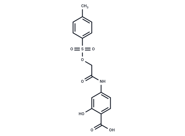
S3I-201 (S3I-201) is a selective Stat3 inhibitor (IC50: 86±33 μM) and low effect towards STAT1/5.

| Pack Size | Price | Availability | Quantity |
|---|---|---|---|
| 5 mg | $50 | In Stock | |
| 10 mg | $63 | In Stock | |
| 25 mg | $98 | In Stock | |
| 50 mg | $151 | In Stock | |
| 100 mg | $280 | In Stock | |
| 200 mg | $421 | In Stock |
| Description | S3I-201 (S3I-201) is a selective Stat3 inhibitor (IC50: 86±33 μM) and low effect towards STAT1/5. |
| Targets&IC50 | STAT3:86 nM (cell free) |
| In vitro | S3I-201 inhibits Stat3.Stat3 complex formation and Stat3 DNA-binding and transcriptional activities. Furthermore, S3I-201 inhibits growth and induces apoptosis preferentially in tumor cells that contain persistently activated Stat3. Constitutively dimerized and active Stat3C and Stat3 SH2 domain rescue tumor cells from S3I-201-induced apoptosis [1]. Preincubation of CD4 T cells from p53(null)CD45.1 mice with various concentrations of S31-201 suppressed IL-6-induced phosphorylation of STAT3 in a dose-dependent manner as early as 15 min after IL-6 addition. Based on the level of suppression of STAT3 phosphorylation, the IC50 of this inhibitor was determined as 38 μM [2]. Although none of these cell lines was sensitive to S3I-201 (S3I-201) alone, S3I-201 potentiated the antiproliferative effect of cetuximab in all three cell lines (HepG2, SK-HEP1, and Huh-7 cells) [3]. |
| In vivo | Compared with control tumors, which continued to grow, human breast tumors in mice that received S3I-201 displayed strong growth inhibition. Strong inhibition of Stat3 DNA-binding activity in residual tumor tissue from mice treated with S3I-201 compared with control tumor [1]. 8- to 10-wk-old p53nullCD45.1 mice were treated with S31-201 3×/wk at 5 mg/kg, using PBS-treated age-matched p53nullCD45.1 mice as controls. STAT3 phosphorylation in the spleen of S31-201-treated mice was markedly reduced as compared with those treated with PBS [2]. S3I-201 attenuated somatotroph tumor growth and GH secretion in a rat xenograft model [4]. |
| Kinase Assay | Briefly, 100 ml of biotinyl-e-Ac-EPQpYEEIEL-OH (in 50 mM Tris/150 mM NaCl, pH 7.5) was added to each well of streptavidin-coated 96-well microtiter plates and incubated with shaking at 4°C overnight. Then plates were rinsed with PBS/Tween 20 and then two times with 200 ml of BSA-T-PBS (0.2% BSA/0.1% Tween 20/PBS). Then 50 ml of Lck-SH2-GST fusion protein (6.4 ng/ml in BSA-T-PBS) was added to each well of the 96-well plate in the presence and absence of 50 ml of S3I-201 (for 30 and 100 mM final concentrations), and the plate was shaken at room temperature for 4 h. After solutions were removed, each well was rinsed four times with BSA-T-PBS (200 ml), and 100 ml of polyclonal rabbit anti-GST antibody (100 ng/ml in BSA-T-PBS) was added to each well and incubated at 4°C overnight. After washing with BSA-T-PBS, 100 ml of 200 ng/ml BSA-T-PBS horseradish peroxidase-conjugated mouse anti-rabbit antibody was added to each well and incubated for 45 min at room temperature. After four washing steps with BSA-T-PBS and three washing steps with PBS-T, 100 ml of peroxidase substrate was added to each well and incubated for 5-15 min. The peroxidase reaction was stopped by adding 100 ml of 1 M sulfuric acid solution, and absorbance was read at 450 nm with an ELISA plate reader [1]. |
| Cell Research | Proliferating cells were treated with or without S3I-201 for up to 48 h. In some cases, cells were first transfected with Stat3C, ST3-NT, or ST3-SH2 domain or mock-transfected for 24 h before treatment with compound for an additional 24–48 h. Cells were then detached and analyzed by annexin V binding according to the manufacturer's protocol and flow cytometry to quantify the percent apoptosis [1]. |
| Animal Research | Six-week-old female athymic nude mice were purchased from Harlan and maintained in the institutional animal facilities approved by the American Association for Accreditation of Laboratory Animal Care. Athymic nude mice were injected in the left flank area s.c. with 5 × 10^6 human breast cancer MDA-MB-231 cells in 100 μl of PBS. After 5–10 days, tumors with a diameter of 3 mm were established. Animals were given S3I-201 i.v. at 5 mg/kg every 2 or 3 days for 2 weeks and monitored every 2 or 3 days. Animals were stratified so that the mean tumor sizes in all treatment were nearly identical. Tumor volume was calculated according to the formula V = 0.52 × a2× b, where a is the smallest superficial diameter and b is the largest superficial diameter [1]. |
| Alias | S3I201, S3I 201 |
| Molecular Weight | 365.36 |
| Formula | C16H15NO7S |
| Cas No. | 501919-59-1 |
| Smiles | Cc1ccc(cc1)S(=O)(=O)OCC(=O)Nc1ccc(C(O)=O)c(O)c1 |
| Relative Density. | 1.507 g/cm3 |
| Storage | Powder: -20°C for 3 years | In solvent: -80°C for 1 year | Shipping with blue ice. | |||||||||||||||||||||||||||||||||||
| Solubility Information | DMSO: 40 mg/mL (109.48 mM), Sonication is recommended. | |||||||||||||||||||||||||||||||||||
Solution Preparation Table | ||||||||||||||||||||||||||||||||||||
DMSO
| ||||||||||||||||||||||||||||||||||||

Copyright © 2015-2025 TargetMol Chemicals Inc. All Rights Reserved.