Shopping Cart
- Remove All
 Your shopping cart is currently empty

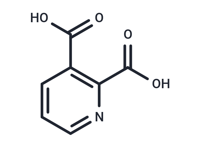
Quinolinic acid (QUIN) is an endogenous N-methyl-D-aspartate (NMDA) receptor agonist synthesized from L-tryptophan via the kynurenine pathway.

| Pack Size | Price | Availability | Quantity |
|---|---|---|---|
| 500 mg | $41 | In Stock | |
| 1 g | $48 | In Stock | |
| 1 mL x 10 mM (in DMSO) | $45 | In Stock |
| Description | Quinolinic acid (QUIN) is an endogenous N-methyl-D-aspartate (NMDA) receptor agonist synthesized from L-tryptophan via the kynurenine pathway. |
| In vitro | QUIN has an uptake system, its neuronal degradation enzyme is rapidly saturated, and the rest of the extracellular QUIN can continue stimulating the NMDA receptor. QUIN (10?μM) prevents of glutamate-induced excitotoxicity in primary cultures of rat cerebellar granule neurons, nevertheless mature organotypic cultures of rat corticostriatal system or caudate nucleus chronically exposed to 100?nM QUIN for up to 7 weeks show focal degeneration characterized by the presence of vacuoles in neuropil, swollen dendrites, occasional swollen post-synaptic elements, and degenerated neurons. In vitro QUIN treatment of human primary fetal neurons leads to a substantial increase of tau phosphorylation at multiple positions. The increase in QUIN-induced phosphorylation of tau is attributed to a decrease in the expression and activity of the major tau phosphatases. QUIN can inhibit B monoamine oxidase (MAO-B) in human brain synaptosomal mitochondria and also can be a potent inhibitor of phosphoenolpyruvate carboxykinase (EC 4.1.1.32) from rat liver cytoplasm, an important enzyme in the gluconeogenesis pathway that converts oxaloacetate to phosphoenolpyruvate. QUIN can increase free radical production by inducing NOS activity in astrocytes and neurons, leading to oxidative stress, increasing both poly(ADP-ribose) polymerase (PARP) activity and extracellular lactate dehydrogenase (LDH) activity [1]. |
| In vivo | Quinolinic acid (QUIN), a neuroactive metabolite of the kynurenine pathway, is normally presented in nanomolar concentrations in human brain and cerebrospinal fluid (CSF) and is often implicated in the pathogenesis of a variety of human neurological diseases. The concentration of QUIN varies among different brain regions, with the cerebral cortex containing approximately 1.8?nmol/g wet weight; almost 2-fold than that found in the hippocampus (1?nmol/g wet weight). Intraarterial administration of either micromolar or millimolar concentrations of QUIN results in only negligible accumulations of this metabolite in the brain, suggesting that the central nervous system (CNS) appears to be well protected by the blood-brain barrier (BBB) from peripheral QUIN. QUIN can also increase glutamate release and inhibit its reuptake by astrocytes, thus increasing its concentration in the microenvironments, causing neurotoxicity and also limiting glutamate to glutamine recycling in astrocytes by decreasing glutamine synthetase activity. Intrastriatal injection of QUIN provokes a decrease in cellular respiration and ATP levels [1]. |
| Alias | QUIN, pyridine-2,3-dicarboxylic acid |
| Molecular Weight | 167.12 |
| Formula | C7H5NO4 |
| Cas No. | 89-00-9 |
| Smiles | OC(=O)C1=C(N=CC=C1)C(O)=O |
| Relative Density. | 1.551 g/cm3 |
| Storage | Powder: -20°C for 3 years | In solvent: -80°C for 1 year | Shipping with blue ice. | |||||||||||||||||||||||||||||||||||
| Solubility Information | DMSO: 50 mg/mL (299.19 mM), Sonication is recommended. | |||||||||||||||||||||||||||||||||||
Solution Preparation Table | ||||||||||||||||||||||||||||||||||||
DMSO
| ||||||||||||||||||||||||||||||||||||

Copyright © 2015-2025 TargetMol Chemicals Inc. All Rights Reserved.