Shopping Cart
- Remove All
 Your shopping cart is currently empty

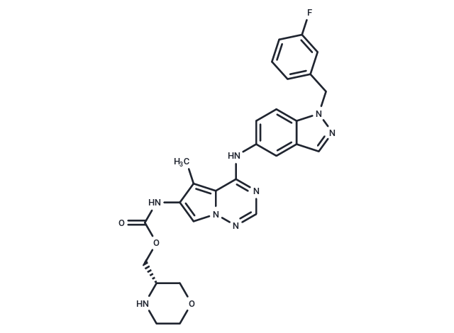
BMS-599626 (AC480) has been used in trials studying the treatment of Cancer, Metastases, and HER2 or EGFR Expressing Advanced Solid Malignancies.

| Pack Size | Price | Availability | Quantity |
|---|---|---|---|
| 10 mg | Inquiry | 35 days | |
| 25 mg | Inquiry | 35 days |
| Description | BMS-599626 (AC480) has been used in trials studying the treatment of Cancer, Metastases, and HER2 or EGFR Expressing Advanced Solid Malignancies. |
| Targets&IC50 | HER2:30 nM, HER4:190 nM, HER1:20 nM |
| In vitro | In vivo administration of BMS-599626, ranging from 60 mg/kg to 240 mg/kg, resulted in a dose-dependent inhibition of Sal2 tumor growth, demonstrating effective antitumor activity in its KPL-4 human breast cancer xenografts. The maximum tolerated dose was identified as 180 mg/kg. Similarly, this compound exhibited comparable antitumor activities in other HER2-amplified xenograft models as well as in various HER1-overexpressing xenograft models. |
| In vivo | MS-599626 effectively inhibits the proliferation of tumor cells expressing high levels of HER1 and/or HER2, including Sal2, BT474, N87, KPL-4, HCC202, HCC1954, HCC1419, AU565, ZR-75-30, MDA-MB-175, GEO, and PC9 cells, with IC50 values of 0.24 μM, 0.31 μM, 0.45 μM, 0.38 μM, 0.94 μM, 0.34 μM, 0.75 μM, 0.63 μM, 0.51 μM, 0.84 μM, 0.90 μM, and 0.34 μM, respectively. BMS-599626 selectively inhibits the enzymatic activity of recombinant HER1 and HER2 kinases, with IC50 values of 20 nM and 30 nM, respectively. It significantly enhances the radiosensitivity of HN-5 cells expressing EGFR and Her2 by promoting cell cycle redistribution and inhibiting DNA repair. Furthermore, BMS-599626 also inhibits HER4, albeit with lower potency, showing an IC50 of 190 nM. However, it does not significantly inhibit the proliferation of the ovarian tumor cell line A2780 and the fibroblast cell line MRC5, which do not express HER1 or HER2. |
| Kinase Assay | Protein kinase assays: The entire cytoplasmic sequences of HER1, HER2, and HER4 are expressed as recombinant proteins in Sf9 insect cells. HER1 and HER4 are expressed as fusion proteins with glutathione-S-transferase and are purified by affinity chromatography on glutathione-S-Sepharose. HER2 is subcloned into the pBlueBac4 vector and expressed as an untagged protein using an internal methionine codon (M687) for translation initiation. The truncated HER2 protein is isolated by chromatography on a column of DEAE-Sepharose equilibrated in a buffer that contains 0.1 M NaCl, and the recombinant protein is eluted with a buffer containing 0.3 M NaCl. For the HER kinase assays, reaction volumes are 50 μL and contains 10 ng of glutathione-S-transferase fusion protein or 150 ng of partially purified HER2. The mixtures also contains 1.5 μM poly(Glu/Tyr) (4:1), 1 μM ATP, 0.15 μCi [γ-33P]ATP, 50 mM Tris-HCl (pH 7.7), 2 mM DTT, 0.1 mg/mL bovine serum albumin, and 10 mM MnCl2. Reactions are allowed to proceed at 27°C for 1 hour and are terminated by the addition of 10 μL of a stop buffer (2.5 mg/mL bovine serum albumin and 0.3 M EDTA), followed by a 108-μL mixture of 3.5 mM ATP and 5% trichloroacetic acid. Acid-insoluble proteins are recovered on GF/C Unifilter plates with a Filtermate harvester. Incorporation of radioactive phosphate into the poly(Glu/Tyr) substrate is determined by liquid scintillation counting. Percent inhibition of kinase activity is determined by nonlinear regression analyses and data are reported as the inhibitory concentration required to achieve 50% inhibition relative to control reactions (IC50). Data are the averages of triplicate determinations. All other tyrosine kinases are also assayed using poly(Glu/Tyr) as a substrate. Kinetics of HER1 and HER2 inhibition are determined in reaction mixtures that contains varying concentrations of ATP and BMS-599626. |
| Cell Research | All cell lines are maintained in RPMI 1640 supplemented with 10% fetal bovine serum, 100 units/mL penicillin, and 100 μg/mL streptomycin. Cells are plated at 1,000 per well in 96-well plates and are cultured for 24 hours before BMS-599626 is added. BMS-599626 is diluted in culture medium such that the final concentrations of DMSO are ≤ 1%. Following the addition of BMS-599626, the cells are cultured for an additional 72 hours before cell viability is determined by measuring the conversion of 3-(4,5-dimethylthiazol-2-yl)-2,5-diphenyltetrazolium bromide dye with the CellTiter96 kit. For some cell lines, there is a lack of a correlation between 3-(4,5-dimethylthiazol-2-yl)-2,5-diphenyltetrazolium bromide dye metabolism and cell number, and a thymidine uptake assay is used to measure proliferation of these cell lines. Cells are plated in 96-well plates and treated with compounds as above. At the end of the 72-hour incubation, cells are pulsed with [3H]thymidine (0.4 μCi/well) for 3 hours before they are harvested. Cells are digested with 2.5% trypsin for 10 minutes at 37 °C and are harvested by filtration using a Packard Filtermate Harvester and GF/C Unifilter plates. Incorporation of radioactive thymidine into nucleic acids is determined by liquid scintillation counting.(Only for Reference) |
| Alias | AC480 |
| Molecular Weight | 530.55 |
| Formula | C27H27FN8O3 |
| Cas No. | 714971-09-2 |
| Smiles | Cc1c(NC(=O)OC[C@@H]2COCCN2)cn2ncnc(Nc3ccc4n(Cc5cccc(F)c5)ncc4c3)c12 |
| Relative Density. | 1.484 g/cm3 |
| Storage | Powder: -20°C for 3 years | In solvent: -80°C for 1 year | Shipping with blue ice. | ||||||||||||||||||||||||||||||||||||||||
| Solubility Information | H2O: <1 mg/mL DMSO: 104 mg/mL (196.02 mM), Sonication is recommended. Ethanol: 16 mg/mL (30.16 mM), Sonication is recommended. | ||||||||||||||||||||||||||||||||||||||||
Solution Preparation Table | |||||||||||||||||||||||||||||||||||||||||
Ethanol/DMSO
DMSO
| |||||||||||||||||||||||||||||||||||||||||

Copyright © 2015-2025 TargetMol Chemicals Inc. All Rights Reserved.