
Shopping Cart
- Remove All
 Your shopping cart is currently empty

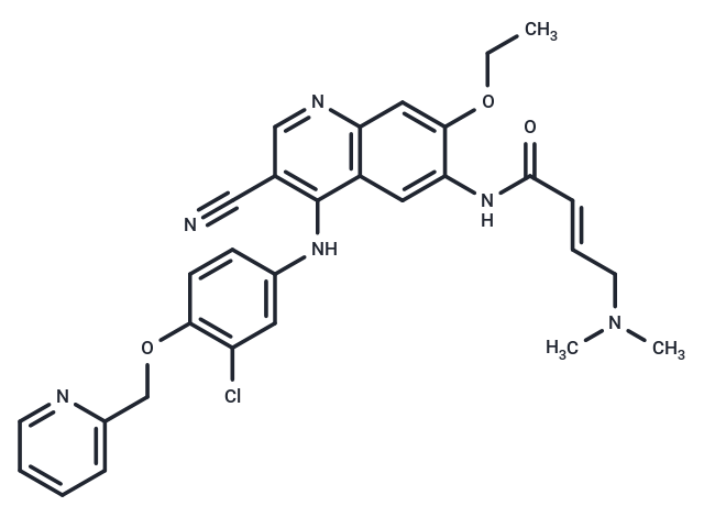
Neratinib (HKI-272) (HKI-272) is an orally available, irreversible tyrosine kinase inhibitor for HER2 and EGFR (IC50: 59/92 nM), respectively.
| Pack Size | Price | Availability | Quantity |
|---|---|---|---|
| 5 mg | $30 | In Stock | |
| 10 mg | $43 | In Stock | |
| 25 mg | $64 | In Stock | |
| 50 mg | $85 | In Stock | |
| 100 mg | $126 | In Stock | |
| 200 mg | $197 | In Stock | |
| 500 mg | $328 | In Stock |
| Description | Neratinib (HKI-272) (HKI-272) is an orally available, irreversible tyrosine kinase inhibitor for HER2 and EGFR (IC50: 59/92 nM), respectively. |
| Targets&IC50 | HER2:59 nM (cell free), EGFR:92 nM (cell free) |
| In vitro | In a cell-free autophosphorylation assay using the recombinant cytoplasmic domain of HER-2, Neratinib (HKI-272) reduced kinase activity by 50% (IC50) at a concentration of 59 nM. It weakly inhibited two other tyrosine kinases tested, KDR and src (IC50: 800 and 1400 nM). HKI-272 repressed the proliferation of a mouse fibroblast cell line (3T3) transfected with the HER-2 oncogene (3T3/neu) (IC50: 3 nM). HKI-272 also inhibited two other HER-2-overexpressing breast cancer cell lines, SK-Br-3, and BT474 (IC50: 2 nM). HKI-272 inhibited proliferation of the epidermal carcinoma cell line, A431, that overexpresses EGFR (IC50: 81 nM) [1]. Ba/F3 cells expressing EGFRvIII mutants are highly sensitive to HKI-272, (IC50: 9.4 nM), as are Ba/F3 cells expressing EGFR-L858R (IC50: 3.5 nM); EGFR-L858R-T790M-expressing cells are also sensitive to HKI-272 (IC50: 180 nM) [2]. |
| In vivo | HKI-272 reduced tumor growth in a dose-dependent manner when administered orally between 20 mg/kg/day (53% inhibition, day 21) and 80 mg/kg/day (98% inhibition). In a second independent test, HKI-272 showed antitumor effects between 10 mg/kg/day (34% inhibition, day 14) and 40 mg/kg/day (98% inhibition). In xenografts of BT474, HKI-272 treatment repressed tumor growth when administered to animals between 10 mg/kg/day (67% inhibition, day 28) and 40 mg/kg/day (93% inhibition). Phosphorylation of HER-2 was inhibited by 84% within 1 h of administration with HKI-272 (40 mg/kg). Inhibition was sustained at 6 h (97%) and decreased to 43% over 24 h [1]. The three HKI-272 (50 mg/kg, p.o.)-treated mice experienced a more substantial tumor response, an average 88% reduction in tumor volume. Decrease of both total EGFRvIII and phospho-EGFR levels was observed after 1 week of HKI-272 treatment [2]. |
| Kinase Assay | Activity of HER-2 and EGFR cytoplasmic domains was measured by an autophosphorylation assay using time-resolved fluorometry. Compounds were prepared as 10 mg/ml stocks in DMSO and diluted in 25 mm HEPES (pH 7.5; 0.002 ng/ml–20 μg/ml). Enzyme [diluted in 100 mm HEPES (pH 7.5) and 50% glycerol] was incubated with inhibitor in 4 mm HEPES (pH 7.5), 0.4 mm MnCl2, 20 μm sodium vanadate, and 0.2 mm DTT for 15 min at room temperature in 96-well ELISA plates. The kinase reaction was initiated by the addition of 40 μm ATP and 20 mm MgCl2 and allowed to proceed for 1 h at room temperature. Plates were washed, and phosphorylation was detected using Europium-labeled anti-phospho-tyrosine antibodies (15 ng/well). After washing and enhancement steps according to the manufacturer's recommendations, signal was detected using a Victor^2 fluorescence reader (excitation wavelength 340 nm, emission wavelength 615 nm). The concentration of compound that inhibited receptor phosphorylation by 50% (IC50) was calculated from inhibition curves [1]. |
| Cell Research | Cells were plated in 96-well tissue culture plates (3T3, 3T3/neu, 5000 cells/well; A431, SK-Br-3, BT474, MDA-MB-435, and SW620, 10,000 cells/well). The following day, dilutions of compound (0.5 ng/ml–5 μg/ml) were added, and cells were cultured for 2 days (6 days for BT474). Cell proliferation was determined using sulforhodamine B, a protein binding dye. Briefly, cells were fixed with 10% trichloroacetic acid and washed extensively with water. Cells were then stained with 0.1% sulforhodamine B and washed in 5% acetic acid. Protein-associated dye was solubilized in 10 mm Tris, and absorbance was measured at 450 nm (Victor^2). Inhibition of cell proliferation was calculated using the formula: percentage of inhibition = 100 ? 100 (Td ? To/Tc ? To), where Td is the absorbance of drug-treated cells, Tc is the absorbance of untreated cells, and To is the absorbance at the time of drug addition. To values were determined by plating cells separately and fixing them at the time of drug addition. The concentration of compound which inhibits cell proliferation by 50% (IC50) was determined from inhibition curves [1]. |
| Animal Research | Athymic female nude mice (5 animals/group) were implanted s.c. with BT474 tumor fragments (~30 mm^3). When tumors reached 200–300 mg, animals were given a single oral dose (40 mg/kg) of HKI-272 in pH 2.0 water. Tumors from control and treated animals were excised at 1, 3, 6, and 24 h and minced. Tumor fragments were suspended in 10 mm Tris (pH 7.5), 5 mm EDTA, 150 mm NaCl, 1% Triton X-100, 1% sodium deoxycholate, 0.1% SDS, 1 mm phenylmethylsulfonyl fluoride, 10 μg/ml pepstatin, 10 μg/ml leupeptin, 10 μg/ml aprotinin, 2 mm sodium vanadate, and 100 mm sodium fluoride and lysed by homogenization on ice with a polytron. After clarification by centrifugation, protein concentration in lysates was estimated using the Bio-Rad DC protein assay. Sixty μg of lysate pooled from each group were analyzed by SDS-PAGE and immunoblotting with phospho-tyrosine-specific antibodies. Pooled extracts were also immunoprecipitated using 4 μg of anti-HER-2 antibodies for 1 h at 4°C. Immune complexes were collected on protein A-agarose, washed, and analyzed by immunoblotting using phospho-tyrosine-specific antibodies. Extracts from individual tumors were analyzed to determine variability between animals [1]. |
| Alias | HKI-272 |
| Molecular Weight | 557.04 |
| Formula | C30H29ClN6O3 |
| Cas No. | 698387-09-6 |
| Smiles | N(C=1C2=C(C=C(OCC)C(NC(/C=C/CN(C)C)=O)=C2)N=CC1C#N)C3=CC(Cl)=C(OCC4=CC=CC=N4)C=C3 |
| Relative Density. | 1.337 g/cm3 |
| Storage | Powder: -20°C for 3 years | In solvent: -80°C for 1 year | Shipping with blue ice. |
| Solubility Information | H2O: < 1 mg/mL (insoluble or slightly soluble) Ethanol: < 1 mg/mL (insoluble or slightly soluble) DMSO: 5 mg/mL (8.9 mM), Sonication is recommended. 10% DMSO+40% PEG300+5% Tween 80+45% Saline: 0.25 mg/mL (0.45 mM), In vivo: Please add the solvents sequentially, clarifying the solution as much as possible before adding the next one. Dissolve by heating and/or sonication if necessary. Working solution is recommended to be prepared and used immediately. |
 For example, your dosage is 10 mg/kg Each animal weighs 20 g, and the dosage volume is 100 μL . A total of 10 animals were administered, and the formula you used is 5%
  DMSO+30% PEG300+5% Tween 80+60% ddH2O. So your working solution concentration is 2 mg/mL。 (mother liquor concentration of 40 mg/mL), if you need to configure a concentration that exceeds the solubility of the product, please contact us first. main solution, add 300 μLPEG300 mix well and clarify, then add 50 more μL Tween 80, mix well and clarify, then add 600 more μLddH2O mix well and clarify
 Hello! How can I help you today?
Copyright © 2015-2025 TargetMol Chemicals Inc. All Rights Reserved.