Shopping Cart
- Remove All
 Your shopping cart is currently empty

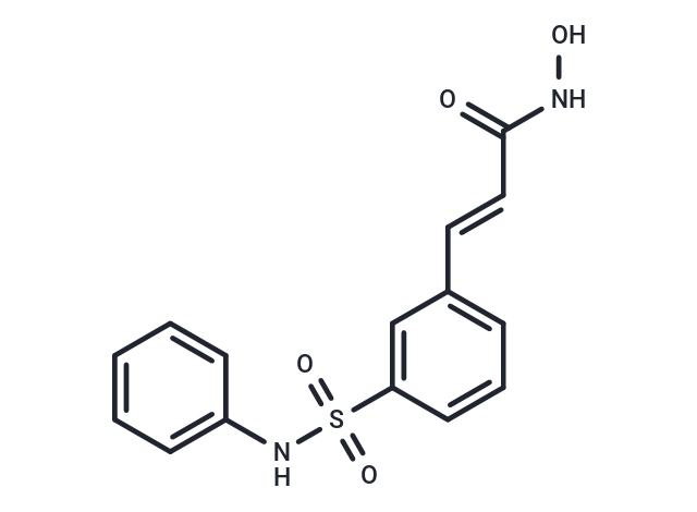
Rac-Belinostat (PX-105684) is a novel hydroxamic acid-type histone deacetylase (HDAC) inhibitor with antineoplastic activity. Belinostat targets HDAC enzymes, thereby inhibiting tumor cell proliferation, inducing apoptosis, promoting cellular differentiation, and inhibiting angiogenesis. This agent may sensitize drug-resistant tumor cells to other antineoplastic agents, possibly through a mechanism involving the down-regulation of thymidylate synthase.

| Pack Size | Price | Availability | Quantity |
|---|---|---|---|
| 10 mg | $37 | In Stock | |
| 25 mg | $59 | In Stock | |
| 50 mg | $88 | In Stock | |
| 100 mg | $130 | In Stock | |
| 200 mg | $208 | In Stock | |
| 1 mL x 10 mM (in DMSO) | $41 | In Stock |
| Description | Rac-Belinostat (PX-105684) is a novel hydroxamic acid-type histone deacetylase (HDAC) inhibitor with antineoplastic activity. Belinostat targets HDAC enzymes, thereby inhibiting tumor cell proliferation, inducing apoptosis, promoting cellular differentiation, and inhibiting angiogenesis. This agent may sensitize drug-resistant tumor cells to other antineoplastic agents, possibly through a mechanism involving the down-regulation of thymidylate synthase. |
| Targets&IC50 | HDAC:27 nM |
| In vitro | In bladder cells of mice, Belinostat induces the expression of p21WAF1, core HDAC, and genes essential for cellular communication. When administered to A2780 and A2780/cp70 xenografts, Belinostat at a dosage of 10 mg/kg significantly delays tumor growth without affecting the body weight of the animals. A higher dose of Belinostat (100 mg/kg) targeting A2780 xenografts alone achieves a tumor inhibition rate of 47%, demonstrating a dose-dependent effect. The combined treatment of Belinostat (100 mg/kg) and Carboplatin (40 mg/kg) extends the delay in tumor growth to 18.6-22.5 days. Moreover, when applied to mice carrying Bortezomib-resistant UMSCC-11A xenografts, the compound exhibits gastrointestinal toxicity. Importantly, the concomitant use of Bortezomib and Belinostat significantly suppresses tumor growth. |
| In vivo | Belinostat exhibits low activity against A2780/cp70 and 2780AD cells, which are derived from A2780 cells resistant to doxorubicin and cisplatin. In ovarian cancer cell lines, Belinostat enhances microtubule acetylation. It inhibits tumor cell growth, including A2780, HCT116, HT29, WIL, CALU-3, MCF7, PC3, and HS852 cells (IC50: 0.2-0.66 μM), through acetylation of histones H3/H4 and PARP cleavage, thereby inducing apoptosis. Belinostat notably suppresses the growth of bladder cancer cells, particularly the 5637 cells, with an accumulation of cells in the G0-G1 phase, a decline in the S phase, and an increase in the G2-M phase. Its inhibitory effect on cell growth is independent of the multidrug-resistant phenotype, although the activity of docetaxel is significantly affected. Belinostat enhances the inhibitory activity of carboplatin or docetaxel on A2780 and OVCAR-3 cells. Within a TGF-β signaling-dependent mechanism, Belinostat activates protein kinase A and reduces survivin mRNA levels. |
| Kinase Assay | Histone Deacetylase Activity: Subconfluent cultures are harvested and washed twice in ice cold PBS and pelleted by centrifugation at 200 × g for 5 min. The cell pellet is resuspended in two volumes of lysis buffer [60 mM Tris buffer (pH 7.4) containing 30% glycerol and 450 mM NaCl] and lysed by three freeze (dry ice) thaw (30 °C water bath) cycles. Cell debris is removed by centrifugation at 1.2 × 10⁴ g for 5 min, and the supernatant is stored at ?80 °C. Histone H4 peptide (sequence SGRGKGGKGLGKGGAKRHRK corresponding to the 20 NH2-terminal residues) is acetylated by a recombinant protein containing the hypoxanthine-aminopterin-thymidine domain of p300, using [3H]acetyl CoA as a source of acetate. H4 peptide (100 μg) is mixed with hypoxanthine-aminopterin-thymidine buffer (50 mM Tris HCl pH 8.0, 5% glycerol, 50 mM KCl, and 0.1 mM EDTA), 1 mM DTT, 1 mM 4-(2-aminoethyl) benzenesulfonylfluoride, 1 × complete protease inhibitors, 50 μL of purified p300, and 1.85 m [3H]acetyl CoA (4.50Ci/mmol) in a final volume of 300 μL and incubated at 30 °C for 45 min. The p300 protein is removed by incubation with 20 μL of 50% Ni-agaroase beads for 1 hour at 4 °C and centrifugation. The supernatant is applied to a 2 mL Sephadex G15 column, and the flow through is collected. One milliliter of distilled Water is gently applied, and three drop fractions are collected; this is repeated until 4–5 mL of distilled Water has been added, and ~40 fractions are collected. Three microliters of each fraction are diluted in 2 mL of scintillation fluid and counted in a scintillation counter to identify the fractions containing the labeled peptide. These fractions are pooled, and 1 μL of the combined sample is measured to assess the radioactivity in every peptide batch (3-7×10³ cpm/μL). For activity assays, the reaction is carried out in a total volume of 150 μL of buffer [60 mM Tris (pH 7.4) containing 30% glycerol] containing 2 μL of cell extract and, where used, 2 μL of belinostat.The reaction is started by the addition of 2 μL of [3H] labeled substrate (acetylated histone H4 peptide corresponding to the 20 NH2-terminal residues). Samples are incubated at 37 °C for 45 min, and the reaction stopped by the addition of HCl and acetic acid (0.72 and 0.12 M final concentrations, respectively). Released [3H]acetate is extracted into 750 μL of ethyl acetate, and samples are centrifuged at 1.2× 10⁴ g for 5 min. The upper phase (600 μL) is transferred to 3 mL of scintillation fluid and counted. |
| Cell Research | Tumor cell lines are seeded in 5 mL of medium at a density of 8 × 10⁴ cells/25 cm2 flask and incubated for 48 hours. Cells are exposed to Belinostat (0.016 to 10 μM) for 24 hours. The medium is removed, and 1 mL of trypsin/EDTA is added to each flask. Once the cells have detached, 1 mL of medium is added, the cells are resuspended, and those from the control untreated flask are counted. Cells are diluted and plated into 6-cm Petri dishes (three per flask) at a density of 0.5-2× 10³ cells/dish depending on the cell line. Cells from the drug-treated flasks are diluted and plated as for the control flasks. Dishes are incubated for 10–15 days at 37 °C. Cells are washed with PBS, fixed in methanol, and stained with crystal violet, and colonies that contained ≥50 cells counted. Sensitivity is expressed as the IC50 defined as the concentration of belinostat required to reduce the number of colonies to 50% of that of the control untreated cells.(Only for Reference) |
| Alias | PXD101, PX-105684, NSC726630 |
| Molecular Weight | 318.35 |
| Formula | C15H14N2O4S |
| Cas No. | 414864-00-9 |
| Smiles | ONC(=O)\C=C\c1cccc(c1)S(=O)(=O)Nc1ccccc1 |
| Relative Density. | 1.427g/cm3 |
| Storage | Powder: -20°C for 3 years | In solvent: -80°C for 1 year | Shipping with blue ice. | |||||||||||||||||||||||||||||||||||
| Solubility Information | Ethanol: < 1 mg/mL (insoluble or slightly soluble) H2O: < 1 mg/mL (insoluble or slightly soluble) DMSO: 65 mg/mL (204.18 mM), Sonication is recommended. | |||||||||||||||||||||||||||||||||||
Solution Preparation Table | ||||||||||||||||||||||||||||||||||||
DMSO
| ||||||||||||||||||||||||||||||||||||

Copyright © 2015-2025 TargetMol Chemicals Inc. All Rights Reserved.