Shopping Cart
- Remove All
 Your shopping cart is currently empty

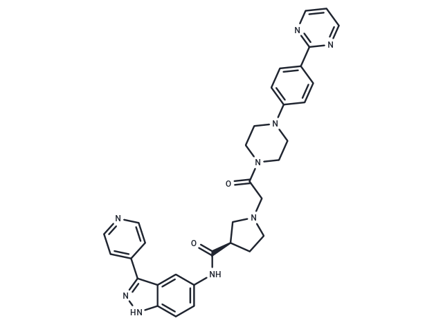
SCH772984 is an ERK inhibitor that inhibits ERK1 and ERK2 (IC50=4/1 nM) and is highly selective and ATP-competitive. SCH772984 exhibits antitumor activity against BRAF or RAS mutant cells.

| Pack Size | Price | Availability | Quantity |
|---|---|---|---|
| 1 mg | $48 | In Stock | |
| 2 mg | $70 | In Stock | |
| 5 mg | $100 | In Stock | |
| 10 mg | $151 | In Stock | |
| 25 mg | $307 | In Stock | |
| 50 mg | $460 | In Stock | |
| 100 mg | $676 | In Stock | |
| 200 mg | $962 | In Stock | |
| 500 mg | $1,430 | In Stock | |
| 1 mL x 10 mM (in DMSO) | $133 | In Stock |
| Description | SCH772984 is an ERK inhibitor that inhibits ERK1 and ERK2 (IC50=4/1 nM) and is highly selective and ATP-competitive. SCH772984 exhibits antitumor activity against BRAF or RAS mutant cells. |
| Targets&IC50 | ERK1:4 nM (cell free), ERK2:1 nM (cell free) |
| In vitro | METHODS: Twenty-one melanoma cell lines containing mutations in the BRAF gene were treated with SCH772984 (0-10 µM) for 72-120 h. Cell viability was measured by CellTiter-Glo Luminescent Cell Viability Assay. RESULTS: Among 21 cell lines, sensitivity to SCH-772984 was categorized into 3 groups: highly sensitive (IC50< 1 µM), moderately sensitive (IC50= 1-2 µM) and resistant (IC50> 2 µM). [1] METHODS: BRAF mutant A375 cells were treated with SCH772984 (0.1-2 µmol/L) for 4 h, and the expression levels of target proteins were detected by Western Blot. RESULTS: Epirubicin significantly increased sub-G cells in G2/M block. [2] |
| In vivo | METHODS: To detect anti-tumor activity in vivo, SCH772984 (25-50 mg/kg) was administered intraperitoneally to Nude mice bearing MiaPaCa xenografts twice daily for 14 days. RESULTS: Tumor regression was observed at both doses, 9% at the 25 mg/kg dose and 36% at the 50 mg/kg dose. [2] |
| Kinase Assay | SCH772984 was tested in 8-point dilution curves in duplicate against purified ERK1 or ERK2. The enzyme was added to the reaction plate and incubated with the compound before adding a solution of substrate peptide and ATP. Fourteen microliters of diluted enzyme (0.3 ng active ERK2 per reaction) was added to each well of a 384-well plate. The plates were gently shaken to mix the reagents and incubated for 45 minutes at room temperature. The reaction was stopped with 60 μL of IMAP Binding Solution (1:2,200 dilutions of IMAP beads in 1× binding buffer). The plates were incubated at room temperature for an additional 0.5 hours to allow complete binding of phosphopeptides to the IMAP beads. Plates were read on the LJL Analyst [1]. |
| Cell Research | For resistant cell line creation, cells were grown in Dulbecco's modified Eagle medium with 10% heat-inactivated FBS media and increasing concentrations of inhibitor (PLX4032, 0.1–10 μmol/L; GSK1120212, 0.01–1 μmol/L) over approximately 4 to 8 months until resistant cells acquired growth properties similar to na?ve parental cells (at their top drug concentrations). For combination resistance, cells were incubated as above but with alternative dose escalation until a top concentration was acquired (PLX4032 10 μmol/L and GSK1120212 1 μmol/L). Stocks and dilutions of PLX4032, GSK1120212, and SCH772984 were made in DMSO solvent. Cell proliferation experiments were carried out in a 96-well format (six replicates), and cells were plated at a density of 4,000 cells per well. At 24 hours after cell seeding, cells were treated with DMSO or a 9-point IC50 dilution (0.001–10 μmol/L) at a final concentration of 1% DMSO for all concentrations. Viability was assayed 5 days after dosing using the ViaLight luminescence kit following the manufacturer's recommendations (n = 6, mean ± SE). For the cell line panel viability assay, cells were treated with SCH772984 for 4 days and assayed by the CellTiterGlo luminescent cell viability assay. For IncuCyte analysis, cells were plated as above in 96-well plates, and image-based cell confluence data were collected every 2 hours during live growth. For engineered resistant lines, cells were infected with lentivirus produced from lentiORF constructs expressing either RFP, KRASG13D, BRAFV600E, truncated BRAFV600E lacking exons 2–8 (Δ2-8), MEK1P124L, MEK1F129L, or constitutively active MEK1DD (S218D+S222D). Cells were selected in blasticidin (20 μg/mL) and used for ViaLight assays as described above [1]. |
| Animal Research | Nude mice were injected subcutaneously with specific cell lines, grown to approximately 100 mm^3, randomized to treatment groups (10 mice/group), and treated intraperitoneally with either SCH772984 or vehicle according to the dosing schedule indicated in the figure legends. Tumor length (L), width (W), and height (H) were measured during and after the treatment periods by a caliper twice weekly on each mouse and then used to calculate tumor volume using the formula (L × W × H)/2. Animal body weights were measured on the same days twice weekly. Data were expressed as mean ± SEM. Upon completion of the experiment, vehicle- and SCH772984-treated tumor biopsies were processed for Western blot analysis [1]. |
| Molecular Weight | 587.67 |
| Formula | C33H33N9O2 |
| Cas No. | 942183-80-4 |
| Smiles | O=C(CN1CC[C@H](C1)C(=O)Nc1ccc2[nH]nc(-c3ccncc3)c2c1)N1CCN(CC1)c1ccc(cc1)-c1ncccn1 |
| Relative Density. | 1.353 g/cm3 at 20℃ |
| Storage | Powder: -20°C for 3 years | In solvent: -80°C for 1 year | Shipping with blue ice. | |||||||||||||||||||||||||
| Solubility Information | H2O: < 1 mg/mL (insoluble or slightly soluble) Ethanol: < 1 mg/mL (insoluble or slightly soluble) DMSO: 18.33 mg/mL (31.2 mM), Sonication is recommended. | |||||||||||||||||||||||||
| In Vivo Formulation | 10% DMSO+90% Saline: 0.54 mg/mL (0.92 mM), Solution. Please add the solvents sequentially, clarifying the solution as much as possible before adding the next one. Dissolve by heating and/or sonication if necessary. Working solution is recommended to be prepared and used immediately. The formulation provided above is for reference purposes only. In vivo formulations may vary and should be modified based on specific experimental conditions. | |||||||||||||||||||||||||
Solution Preparation Table | ||||||||||||||||||||||||||
DMSO
| ||||||||||||||||||||||||||

Copyright © 2015-2025 TargetMol Chemicals Inc. All Rights Reserved.