Shopping Cart
- Remove All
 Your shopping cart is currently empty

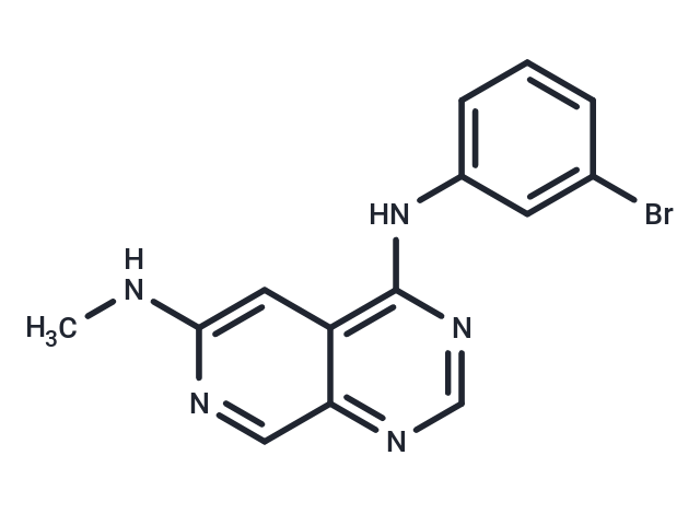
PD 158780 reversibly inhibits auto and transphosphorylation of all four members of the ErbB receptor superfamily: EGFR, ErbB2, ErbB3, and ErbB4 (IC50s: 8μM, 49 nM, 52 nM, and 52 nM in cell assay).

| Pack Size | Price | Availability | Quantity |
|---|---|---|---|
| 1 mg | $34 | In Stock | |
| 5 mg | $77 | In Stock | |
| 10 mg | $122 | In Stock | |
| 25 mg | $228 | In Stock | |
| 50 mg | $397 | In Stock | |
| 100 mg | $582 | In Stock | |
| 1 mL x 10 mM (in DMSO) | $98 | In Stock |
| Description | PD 158780 reversibly inhibits auto and transphosphorylation of all four members of the ErbB receptor superfamily: EGFR, ErbB2, ErbB3, and ErbB4 (IC50s: 8μM, 49 nM, 52 nM, and 52 nM in cell assay). |
| Targets&IC50 | EGFR:0.008 nM (cell free), EGFR:8 μM (Cell Assay), ERB4:52 nM (Cell Assay), ErbB2:49 nM (Cell Assay), ERB3:52 nM (Cell Assay) |
| In vitro | PD158780 inhibited EGF receptor autophosphorylation in A431 human epidermoid carcinoma with IC50 values of 13 nM. PD 158780 was highly specific for the EGF receptor in Swiss 3T3 fibroblasts, inhibiting EGF-dependent receptor autophosphorylation and thymidine incorporation at low nanomolar concentrations. PD 158780 inhibited heregulin-stimulated phosphorylation in the SK-BR-3 and MDA-MB-453 breast carcinomas with IC50 values of 49 and 52 nM, respectively [1]. |
| Kinase Assay | Epidermal growth factor receptor was prepared from human A431 carcinoma cell shed membrane vesicles by immunoaffinity chromatography as previously described, and the assays were carried out as reported previously. The substrate used was based on a portion of phospholipase Cγ1, having the sequence Lys-His-Lys-Lys-LeuAla-Glu-Gly-Ser-Ala-Tyr472-Glu-Glu-Val. The reaction was allowed to proceed for 10 min at room temperature and then was stopped by the addition of 2 mL of 75 mM phosphoric acid. The solution was then passed through a 2.5 cm phosphocellulose disk which bound the peptide. This filter was washed with 75 mM phosphoric acid (5×), and the incorporated label was assessed by scintillation counting in an aqueous fluor. Control activity (no drug) gave a count of approximately 100 000 cpm. At least two independent dose-response curves were done and the IC50 values computed. The reported values are averages; variation was generally ±15% [1]. |
| Cell Research | All cell lines were maintained as monolayers in dMEM/F12, 50:50 containing 10% fetal bovine serum. For growth inhibition assays, dilutions of the designated compound in 10 μL were placed in 24-well Linbro plates (1.7 x 1.6 cm, flat bottom) followed by the addition of cells (2 × 10^4) in 2 mL of medium. The plates were incubated for 72 hr at 37 °C in a humidified atmosphere containing 5% CO 2 in air. Cell growth was determined by counting cells with a Coulter model AM electronic cell counter. For clone formation in soft agar, cells were trypsinized, and 10,000 cells/mL were seeded into DMEM/F12 medium containing 10% fetal bovine serum, 0.4% agarose, and the designated concentration of compound. One milliliter of this solution was placed over a bottom layer of the same medium containing 0.8% agarose in a 35-mm Petri dish and incubated at 37 °C in a humidified atmosphere containing 5% CO 2 in air. After 3 weeks, colonies were stained with p-iodonitrotetrazolium violet (INT) and quantitated with an image analyzer using the software NIH Image version 1.55. Incorporation of radiolabeled thymidine into cellular DNA was monitored by exposing compound-treated or control cells to [methyl)H]thymidine at a concentration of 1 μM and specific activity of 1 μCi/nmol. After 2 hr the cells were trypsinized and injected into 2 mL of ice-cold 15% trichloroacetic acid (TCA). The resulting precipitate was collected on glass fiber filters, washed five times with 2-mL aliquots of ice-cold 15% TCA, dried, and placed in scintillation vials plus 10 mL Ready gel [1]. |
| Molecular Weight | 330.18 |
| Formula | C14H12BrN5 |
| Cas No. | 171179-06-9 |
| Smiles | CNc1cc2c(Nc3cccc(Br)c3)ncnc2cn1 |
| Relative Density. | 1.611g/cm3 |
| Storage | Powder: -20°C for 3 years | In solvent: -80°C for 1 year | Shipping with blue ice. | |||||||||||||||||||||||||||||||||||
| Solubility Information | Ethanol: 8 mg/mL (24.23 mM) DMSO: 30 mg/mL (90.86 mM) | |||||||||||||||||||||||||||||||||||
Solution Preparation Table | ||||||||||||||||||||||||||||||||||||
Ethanol/DMSO
DMSO
| ||||||||||||||||||||||||||||||||||||

Copyright © 2015-2024 TargetMol Chemicals Inc. All Rights Reserved.