Shopping Cart
- Remove All
 Your shopping cart is currently empty

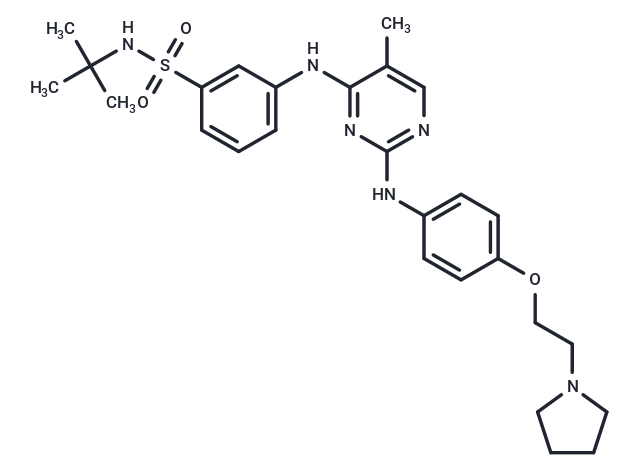
Fedratinib (TG-101348) (TG101348) is an ATP-competitive inhibitor of JAK2 (IC50: 3 nM) with significantly less potent activity against JAK3.

| Pack Size | Price | Availability | Quantity |
|---|---|---|---|
| 5 mg | $45 | In Stock | |
| 10 mg | $63 | In Stock | |
| 50 mg | $115 | In Stock | |
| 100 mg | $136 | In Stock | |
| 200 mg | $228 | In Stock | |
| 500 mg | $497 | In Stock | |
| 1 g | $658 | In Stock | |
| 1 mL x 10 mM (in DMSO) | $52 | In Stock |
| Description | Fedratinib (TG-101348) (TG101348) is an ATP-competitive inhibitor of JAK2 (IC50: 3 nM) with significantly less potent activity against JAK3. |
| Targets&IC50 | JAK2:3 nM (cell free), FLT3:15 nM (cell free), JAK2 (V617F):3 nM (cell free), RET:48 nM (cell free) |
| In vitro | Fedratinib (TG101348) inhibited proliferation of a human erythroblast leukemia (HEL) cell line that harbors the JAK2V617F mutation, as well as a murine pro-B cell line expressing human JAK2V617F (Ba/F3 JAK2V617F), with an IC50 value of approximately 300 nM for either line. Exposure of these cells to TG101348 reduced STAT5 phosphorylation at concentrations that parallel the concentrations required to inhibit cell proliferation [1]. TG101348 inhibited the proliferation of HMC-1.1 (KITV560G) cells, with somewhat lower potency than HMC-1.2 (KITD816V, KITV560G) cells, with IC50's of 740 and 407 nM, respectively. TG101348 did not inhibit phosphorylation of KITV560G or KITD816V within the context of the two HMC-1 clones at concentrations up to 25 μM. TG101348 potently inhibited JAK-STAT signaling in HMC-1.2 cells, the IC50 for JAK2 phosphorylation was 150 and 600 nM; the IC50 for STAT-5 and STAT-3 phosphorylation was ~600 nM [2]. |
| In vivo | During the time course of the study, six animals died in the placebo group, and one animal in the 60 mg/kg drug group at day 18, whereas all animals treated with 120 mg/kg of TG101348 were all alive at study endpoint. There was a 2-fold decrease in JAK2V617F-positive CD71-single-positive early erythroid precursors in the bone marrow of animals at the 120 mg/kg dose compared with vehicle [1]. The maximum tolerated dose was 680 mg/d, and dose-limiting toxicity was a reversible and asymptomatic increase in the serum amylase level. Forty-three patients (73%) continued treatment beyond six cycles; the median cumulative exposure to TG101348 was 380 days [3]. |
| Kinase Assay | IC50 values for TG101348 are determined commercially using the InVitrogen kinase profiling service for a 223 kinase screen that included JAK2 and JAK2V617F or Carna Biosciences for the screen of all Janus kinase family members including JAK1 and Tyk2. ATP concentration is set to approximately the Km value for each kinase [1]. |
| Cell Research | Cells were treated with DMSO and increasing concentrations of inhibitor for 4 hr in RPMI-1640 before collected in 13 Cell Lysis Buffer, containing 1 mM PMSF, and protease inhibitor cocktail tablets. Protein lysates were quantified with BCA assay. Similar protein amounts were mixed with Laemmli sample buffer plus b-mercaptoethanol, boiled for 5 min, and separated on a 4%–15% Tris-HCl gradient electrophoresis gel. Gels were blotted onto a 0.45 mm nitrocellulose membrane, which was blocked in 5% nonfat dry milk and incubated with primary antibodies in either blocking solution or 5% BSA. The membranes were subsequently incubated with a mixture of donkey anti-rabbit IgG conjugated with infrared fluorophore (700 nm emission) and goat anti-mouse IgG conjugated with infrared fluorophore (800 nm emission). Following washing with PBS, the membranes were scanned on an Odyssey scanner to detect total (red) and phospho-STAT5 (green) proteins [1]. |
| Animal Research | The murine BM transplant model was generated and analyzed exactly as previously described. Briefly, C57BL/6 mice were intravenously injected with 1×10^6 whole bone marrow expressing JAK2V617F. Full development of disease was assessed with differential peripheral blood counts at day 26 after bone marrow transplantation. TG101348 was administered by oral gavage twice daily (b.i.d.) at 60 mg/kg, 120 mg/kg, or placebo from day 28 on for 42 days. Differential blood counts were assessed by retro-orbital nonlethal eye bleeds using EDTA glass capillary tubes before study initiation, during the study, and at study endpoints. C57/Bl6 mice were sacrificed at study endpoint or at times indicated based on an IUCAC-approved protocol that includes assessment of morbidity by > 10% loss of weight, scruffy appearance, lethargy, and/or splenomegaly extending across the midline. For histopathology, tissues were fixed in 10% neutral buffered formalin, embedded in paraffin, and stained with hematoxylin and eosin or, to assess for fibrosis, stained with reticulin. Images of histological slides were obtained on a Nikon Eclipse E400 microscope equipped with a SPOT RT color digital camera model 2.1.1. Images were analyzed in Adobe Photoshop 6.0. For flow cytometry, cells were washed in PBS, washed in 2% fetal bovine serum, blocked with Fc-Block for 10 min on ice, and stained with monoclonal antibodies in PBS and 2% FCS for 30 min on ice. Antibodies used were allophycocyanin (APC)-conjugated ter119, Gr-1, CD4, and B220 and phycoerythrin (PE)-conjugated, Mac1, CD8 (all 1:200), and CD71(1:100) rat anti-mouse. After washing, cells were resuspended in PBS and 2% FCS containing 0.5 mg/ml 7-amino-actinomycin D (7-AAD) to allow discrimination of nonviable cells. Flow cytometry was performed on a FACS Calibur cytometer, at least 10,000 events were acquired, and data were analyzed using FloJo software.The results are presented as graphs and representative dot plots of viable cells selected on the basis of scatter and 7-AAD staining [1]. |
| Alias | TG-101348, SAR 302503 |
| Molecular Weight | 524.68 |
| Formula | C27H36N6O3S |
| Cas No. | 936091-26-8 |
| Smiles | Cc1cnc(Nc2ccc(OCCN3CCCC3)cc2)nc1Nc1cccc(c1)S(=O)(=O)NC(C)(C)C |
| Relative Density. | 1.247g/cm3 |
| Storage | Powder: -20°C for 3 years | In solvent: -80°C for 1 year | Shipping with blue ice. | |||||||||||||||
| Solubility Information | 10% DMSO+40% PEG300+5% Tween 80+45% Saline: 9.3 mg/mL (17.73 mM), In vivo: Please add the solvents sequentially, clarifying the solution as much as possible before adding the next one. Dissolve by heating and/or sonication if necessary. Working solution is recommended to be prepared and used immediately. DMSO: 50 mg/mL (95.3 mM), Sonication is recommended. H2O: < 1 mg/mL (insoluble or slightly soluble) Ethanol: < 1 mg/mL (insoluble or slightly soluble) | |||||||||||||||
Solution Preparation Table | ||||||||||||||||
DMSO
| ||||||||||||||||

Copyright © 2015-2025 TargetMol Chemicals Inc. All Rights Reserved.