Shopping Cart
- Remove All
 Your shopping cart is currently empty

BCI-215 causes selective cancer cell cytotoxicity in part through non-redox-mediated activation of MAPK signaling

| Pack Size | Price | Availability | Quantity |
|---|---|---|---|
| 1 mg | $42 | In Stock | |
| 5 mg | $98 | In Stock | |
| 10 mg | $152 | In Stock | |
| 25 mg | $289 | In Stock | |
| 50 mg | $455 | In Stock | |
| 100 mg | $662 | In Stock |
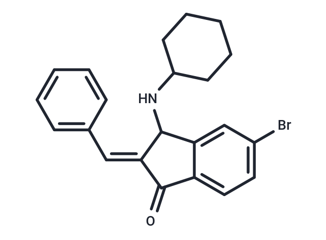
| Description | BCI-215 causes selective cancer cell cytotoxicity in part through non-redox-mediated activation of MAPK signaling |
| In vitro | In MDA-MB-231 human breast cancer cells, BCI-215 inhibited cell motility, caused apoptosis but not primary necrosis, and sensitized cells to lymphokine-activated killer cell activity. Mechanistically, BCI-215 induced rapid and sustained phosphorylation of extracellular signal-regulated kinase (ERK), p38, and c-Jun N-terminal kinase (JNK) in the absence of reactive oxygen species, and its toxicity was partially rescued by inhibition of p38 but not JNK or ERK. BCI-215 also hyperactivated MKK4/SEK1, suggesting activation of stress responses[1]. |
| Cell Research | Peripheral blood mononuclear cells were obtained from healthy volunteers. Cells were cultured in RPMI 1640 supplemented with 10% fetal calf serum, 1% glutamine, and 1% penicillin/streptomycin, and stimulated with 6,000 IU of Interleukin 2 for 24 hours. After incubation, cells were washed with PBS and counted. In parallel, MDA-MB-231 cells were pretreated in a 384-well plate with vehicle or BCI-215 (3 μM). After 24 hours in culture, medium was replaced and peripheral blood mononuclear cells (PBMCs) added in 2-fold serial dilutions starting with a 50-fold excess of PBMCs in triplicate. After 24 hours of coculture, cells were fixed with formaldehyde/Hoechst 33342, washed twice with PBS, and imaged on the ArrayScan II. Cancer cells were identified by their larger nuclei compared with PBMCs, setting a size gate in the Hoechst channel. In experiments with chemotherapeutics, cells carrying a biosensor consisting of a mitochondrial targeting sequence derived from cytochrome c oxidase VIII linked to GFP that is a surrogate for cytochrome c release from mitochondria were pretreated for 24 hours with cisplatin (2 μM) or doxorubicin (400 nM), exposed to LAK, and cancer cells were identified and quantified by green fluorescence. Cell densities were normalized to those in the absence of PBMCs. Mean cell densities from multiple independent experiments were averaged and plotted in GraphPad Prism version 7.00[1]. |
| Molecular Weight | 396.32 |
| Formula | C22H22BrNO |
| Cas No. | 1245792-67-9 |
| Smiles | Brc1ccc2C(=O)\C(=C\c3ccccc3)C(NC3CCCCC3)c2c1 |
| Relative Density. | 1.37 g/cm3 (Predicted) |
| Storage | Powder: -20°C for 3 years | In solvent: -80°C for 1 year | Shipping with blue ice. | ||||||||||||||||||||
| Solubility Information | DMSO: 3.96 mg/mL (10 mM), Sonication is recommended. | ||||||||||||||||||||
Solution Preparation Table | |||||||||||||||||||||
DMSO
| |||||||||||||||||||||

Copyright © 2015-2025 TargetMol Chemicals Inc. All Rights Reserved.