Shopping Cart
- Remove All
 Your shopping cart is currently empty

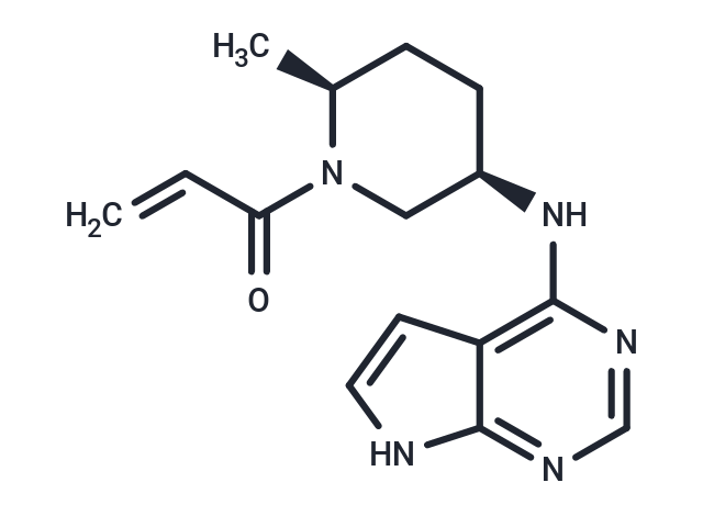
Ritlecitinib (PF-06651600) is an orally available, selective JAK3 inhibitor with an IC50 of 33.1 nM and does not affect the activity of JAK1/2.

| Pack Size | Price | Availability | Quantity |
|---|---|---|---|
| 2 mg | $39 | In Stock | |
| 5 mg | $64 | In Stock | |
| 10 mg | $113 | In Stock | |
| 25 mg | $228 | In Stock | |
| 50 mg | $347 | In Stock | |
| 100 mg | $522 | In Stock | |
| 200 mg | $743 | In Stock | |
| 500 mg | $1,120 | In Stock | |
| 1 mL x 10 mM (in DMSO) | $70 | In Stock |
| Description | Ritlecitinib (PF-06651600) is an orally available, selective JAK3 inhibitor with an IC50 of 33.1 nM and does not affect the activity of JAK1/2. |
| Targets&IC50 | JAK3:33.1 nM (cell free) |
| In vitro | METHODS: The effects of Ritlecitinib on the activity of JAK1/2/3 kinases were investigated in vitro in the presence of physiologically relevant ATP concentrations (1 mM). RESULTS Ritlecitinib inhibited JAK3 kinase activity with an IC50 of 33.1 nM, but had no activity against JAK1, JAK2, and TYK2 (IC50 > 10000 nM). [1] METHODS: The potency and selectivity of F-06651600 in human whole blood total lymphocytes were evaluated by flow cytometry (FACS). RESULTS The IC50 values of Ritlecitinib for inhibiting IL-2, IL-4, IL-7, and IL-15-induced STAT5 phosphorylation were 244, 340, 407, and 266 nM, respectively; the IC50 value of Ritlecitinib for inhibiting IL-21-induced STAT3 phosphorylation was 355 nM. [1] |
| In vivo | METHODS: Ritlecitinib (PF-06651600) (3, 10, 30 mg/kg, oral, once daily) was used to treat two rodent models of arthritis and encephalomyelitis in mice, and its effects were studied. RESULTS Ritlecitinib treatment significantly reduced disease severity in mouse plantar swelling as measured by plethysmography; in a rat adjuvant arthritis (AIA) model, Ritlecitinib reduced plantar swelling with an unbound EC50 of 169 nM. [1] |
| Kinase Assay | His-tagged recombinant human TYK2 kinase domain was expressed in SF21/baculovirus and purified using a two-step affinity (Ni-NTA) and size-exclusion (SEC S200) purification method.Test compounds were solubilized in DMSO to a stock concentration of 30 mM.Compounds were diluted in DMSO to create an 11-point half log dilution series with a top concentration of 600 μM.The test compound plate also contained positive control wells containing a known inhibitor to define 100% inhibition and negative control wells containing DMSO to define no inhibition.The compound plates were diluted 1 to 60 in the assay,resulting in a final assay compound concentration range of 10 μM to 100 pM and a final assay concentration of 1.7% DMSO.Test compounds and controls solubilized in 100% DMSO were added (250 nL) to a 384 well polypropylene plate (Matrical) using a non contact acoustic dispenser.Kinase assays were carried out at room temperature in a 15 μL reaction buffer containing 20 mM HEPES,pH 7.4,10 mM magnesium chloride,0.01% bovine serum albumin (BSA),0.0005% Tween 20 and 1mM Dithiothreitol (DTT).Reaction mixtures contained 1 μM of a fluorescently labeled synthetic peptide,at a concentration less than the apparent Michaelis-Menten constant (Km) (5FAM-KKSRGDYMTMQID for JAK1 and TYK2 and FITC-KGGEEEEYFELVKK for JAK2 and JAK3).Reaction mixtures contained adenosine triphosphate (ATP) at either a level equal to the apparent Km for ATP (40 μM for JAK1,4 μM for JAK2,4 μM for JAK3 and 12 μM for TYK2) or at 1 mM ATP.Compound was added to the buffer containing ATP and substrate and immediately after this step the enzyme was added to begin the reaction.The assays were stopped with 15 μL of a buffer containing 180 mM HEPES,pH=7.4,20 mM EDTA,0.2% Coating Reagent,resulting in a final concentration of 10 mM EDTA,0.1% Coating Reagent and 100 mM HEPES,pH=7.4. |
| Cell Research | Human CD4+ T cells were purified from buffy coat with RosetteSep CD4+ T Cell Enrichment Cocktail and skewed for 6 days with cytokine cocktails (25 ng/mL of IL-6, 25 ng/mL of IL-23, 12.5 ng/mL of IL-1β, 25 ng/mL of IL21, 5 ng/mL of TGFβ1, 10 μg/ml of anti-CD3 antibody (pre-coated on plate surface) and 1 μg/mL of anti-CD28 antibody) in the presence of JAK inhibitors at 10 different concentrations. Supernatants were harvested and the concentrations of IL-17A were determined with MSD assay following the protocol provided by the manufacturer. To study the effect of PF-06651600 on Th17 cells post-differentiation, skewed Th17 cells were washed, rested with X-VIVO 15 medium for overnight and resuspended in medium containing the same concentrations of cytokines as during skewing but without anti-CD3 or anti-CD28 antibodies, in the presence of PF-06651600 at 10 different concentrations for 2 additional days. On Day 9, supernatant was harvested from each well and IL-17A was determined as described above [1]. |
| Animal Research | The effect of JAK3 inhibition by PF-06651600 was evaluated in vivo using a therapeutic dosing paradigm in a rat adjuvant-induced arthritis. The efficacy of this molecule was evaluated in three separate studies using successively lower doses. Arthritis was induced by immunization of female Lewis rats (8 to 10 weeks old) via intradermal injection at the base of the tail with complete Freund's adjuvant with three 50 μL injections (15 mg/mL Mycobacterium tuberculosis) in incomplete Freund's adjuvant. Seven days after the initial immunization, the baseline hind paw volume of the immunized rats was measured via plethysmograph. The rats were monitored daily for signs of arthritis including change in body weight and hind paw volume measurement. When individual hind paw volume measurements indicated an increase of 0.2 mL (or greater) in a single hind paw, animals were randomly assigned to a treatment group. Daily treatment with PF-06651600 was administered via oral gavage. Treatment groups for Experiment 1 were: 80, 15, or 6 mg/kg or vehicle (2% Tween 80 /0.5% methylcellulose/deionized water). Treatment groups for Experiment 2 were: 30, 10, and 3 mg/kg or vehicle (0.5% methylcellulose / de-ionized water/ 1 mEQ hydrochloric acid). Treatment groups for Experiment 3 were: 10, 1, 0.3 and 0.1 mg/kg or vehicle (0.5% methylcellulose/de-ionized water/ 1 mEQ hydrochloric acid). Dosing began once individuals were enrolled into respective groups. Treatment continued for 7 days. At the conclusion of the study, whole blood was taken at 15 minutes post-dose (peak concentration in plasma) for analysis of STAT phosphorylation, and plasma was taken for exposure concentration in PF-06651600 dosed groups [1]. |
| Alias | PF-06651600 |
| Molecular Weight | 285.34 |
| Formula | C15H19N5O |
| Cas No. | 1792180-81-4 |
| Smiles | C[C@H]1CC[C@H](CN1C(=O)C=C)Nc1ncnc2[nH]ccc12 |
| Relative Density. | 1.272 g/cm3 (Predicted) |
| Storage | Powder: -20°C for 3 years | In solvent: -80°C for 1 year | Shipping with blue ice. | |||||||||||||||||||||||||||||||||||
| Solubility Information | DMSO: 130 mg/mL (455.6 mM), Sonication is recommended. | |||||||||||||||||||||||||||||||||||
Solution Preparation Table | ||||||||||||||||||||||||||||||||||||
DMSO
| ||||||||||||||||||||||||||||||||||||

Copyright © 2015-2025 TargetMol Chemicals Inc. All Rights Reserved.