Shopping Cart
- Remove All
 Your shopping cart is currently empty

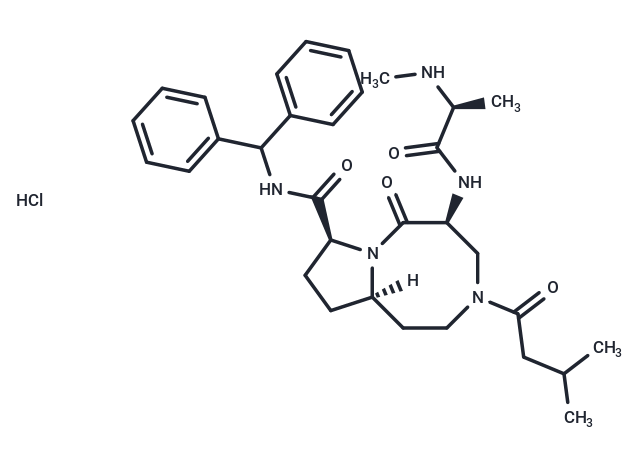
Xevinapant hydrochloride (AT-406 HCl) , an orally active antagonist of multiple inhibitor of apoptosis proteins(IAP), inhibits progression of human ovarian Y binding to XIAP-BIR3, cIAP1-BIR3 and cIAP2-BIR3 with Ki of 66.4 nM, 1.9 nM, and 5.1 nM, 50- to 100-fold higher affinities than the Smac AVPI peptide.

| Pack Size | Price | Availability | Quantity |
|---|---|---|---|
| 1 mg | $35 | In Stock |
| Description | Xevinapant hydrochloride (AT-406 HCl) , an orally active antagonist of multiple inhibitor of apoptosis proteins(IAP), inhibits progression of human ovarian Y binding to XIAP-BIR3, cIAP1-BIR3 and cIAP2-BIR3 with Ki of 66.4 nM, 1.9 nM, and 5.1 nM, 50- to 100-fold higher affinities than the Smac AVPI peptide. |
| Targets&IC50 | XIAP BIR3:66.4 nM(Ki), CIAP1-BIR3:1.9 nM(Ki), CIAP2-BIR3:5.1 nM(Ki) |
| In vitro | AT-406 is a Smac mimetic and appears to mimic closely the AVPI peptide in both hydrogen bonding and hydrophobic interactions with XIAP, with additional hydrophobic contacts with W323 of XIAP. AT-406 is more sensitive to these IAPs than Smac AVPI peptide with 50-100 fold binding affinities. AT-406 (at 1 μM) completely restores the activity of caspase-9, which is suppressed by 500 nM XIAP BIR3 in a cell-free system. In MDA-MB-231 cell, AT-406 induces rapid cellular cIAP1 degradation and also pulls down the cellular XIAP protein. AT-406 effectively inhibits lots of human cancer cell lines and shows IC50 of 144 and 142 nM in MDA-MB-231 cell and SK-OV-3 ovarian cell, with low toxicity against normal-like human breast epithelial MCF-12F cells and primary human normal prostate epithelial cells. AT-406 induces apoptosis in MDA-MB-231 cell by inducing activation of caspase-3 and cleavage of PARP. [1] |
| Kinase Assay | Fluorescence Polarization Based Assays for XIAP, cIAP1, and cIAP2 BIR3 Proteins: FL-AT-406 (the fluorescently tagged AT-406) is employed to develop a set of new FP assays for determination of the binding affinities of Smac mimetics to XIAP, cIAP-1, and cIAP-2 BIR3 proteins. The Kd value of FL-AT-406 to each IAP protein is determined by titration experiments using a fixed concentration of FL-AT-406 and different concentrations of the protein up to full saturation. Fluorescence polarization values are measured using an Infinite M-1000 plate reader in Microfluor 2 96-well, black, round-bottom plates. To each well, FL-AT-406 (2, 1, and 1 nM for experiments with XIAP BIR3, cIAP-1 BIR3, and cIAP-2 BIR3, respectively) and different concentrations of the protein are added to a final volume of 125 μL in the assay buffer (100 mM potassium phosphate, pH 7.5, 100 μg/mL bovine γ-globulin, 0.02% sodium azide, with 4% DMSO). Plates are mixed and incubated at room temperature for 2-3 hours with gentle shaking. The polarization values in millipolarization units (mP) are measured at an excitation wavelength of 485 nm and an emission wavelength of 530 nm. Equilibrium dissociation constants (Kd) are then calculated by fitting the sigmoidal dose-dependent FP increases as a function of protein concentrations using Graphpad Prism 5.0 software. In competitive binding experiments for XIAP3 BIR3, AT-406 is incubated with 20 nM XIAP BIR3 protein and 2 nM FL-AT-406 in the assay buffer (100 mM potassium phosphate, pH 7.5; 100 μg/mL bovine γ-globulin; 0.02% sodium azide). In competitive binding experiments for cIAP1 BIR3 protein, 3 nM protein and 1 nM FL-AT-406 are used. In competitive binding experiments for cIAP2 BIR3, 5 nM protein and 1 nM FL-AT-406 are used. For each competitive binding experiment, polarization values are measured after 2-3 hours of incubation using an Infinite M-1000 plate reader.The IC50 value, the inhibitor concentration at which 50% of the bound tracer is displaced, is determined from the plot using nonlinear least-squares analysis. Curve fitting is performed using the PRISM software. A Ki value for AT-406 is calculated. |
| Cell Research | Cells are seeded in 96-well flat bottom cell culture plates at a density of (3-4) × 103 cells/well with AT-406 and incubated for 4 days. The rate of cell growth inhibition after treatment with different concentrations of AT-406 is determined by assaying with (2-(2-methoxy-4-nitrophenyl)-3-(4-nitrophenyl)-5-(2,4-disulfophenyl)-2H-tetrazolium monosodium salt (WST-8). WST-8 is added to each well to a final concentration of 10%, and then the plates are incubated at 37 °C for 2−3 hours. The absorbance of the samples is measured at 450 nm using a TECAN ULTRA reader. Concentration of AT-406 that inhibited cell growth by 50% (IC50) is calculated by comparing absorbance in the untreated cells and the cells treated with AT-406. (Only for Reference) |
| Alias | SM-406, AT-406 HCl |
| Molecular Weight | 598.19 |
| Formula | C32H44ClN5O4 |
| Cas No. | 1071992-57-8 |
| Smiles | Cl.[H][C@]12CC[C@H](N1C(=O)[C@H](CN(CC2)C(=O)CC(C)C)NC(=O)[C@H](C)NC)C(=O)NC(c1ccccc1)c1ccccc1 |
| Relative Density. | no data available |
| Storage | Powder: -20°C for 3 years | In solvent: -80°C for 1 year | Shipping with blue ice. | |||||||||||||||||||||||||||||||||||
| Solubility Information | DMSO: 100 mg/mL (167.17 mM), Sonication is recommended. Ethanol: 100 mg/mL (167.17 mM), Sonication is recommended. H2O: <1 mg/mL | |||||||||||||||||||||||||||||||||||
Solution Preparation Table | ||||||||||||||||||||||||||||||||||||
DMSO/Ethanol
| ||||||||||||||||||||||||||||||||||||

Copyright © 2015-2025 TargetMol Chemicals Inc. All Rights Reserved.