Shopping Cart
- Remove All
 Your shopping cart is currently empty

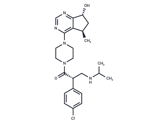
Ipatasertib (GDC-0068) is a selective, ATP-competitive pan-Akt inhibitor that inhibits Akt1 (IC50:5 nM), Akt2 (IC50:18 nM), and Akt3 (IC50:8 nM). Ipatasertib (GDC-0068) can lead to p53-independent PUMA activation by inhibiting Akt, thereby activating FoxO3a and NF-κB simultaneously, directly binding to the PUMA promoter, upregulating PUMA transcription and Bax-mediated intrinsic mitochondrial apoptosis.

| Pack Size | Price | Availability | Quantity |
|---|---|---|---|
| 2 mg | $39 | In Stock | |
| 5 mg | $55 | In Stock | |
| 10 mg | $80 | In Stock | |
| 25 mg | $143 | In Stock | |
| 50 mg | $239 | In Stock | |
| 100 mg | $369 | In Stock | |
| 1 mL x 10 mM (in DMSO) | $56 | In Stock |
| Description | Ipatasertib (GDC-0068) is a selective, ATP-competitive pan-Akt inhibitor that inhibits Akt1 (IC50:5 nM), Akt2 (IC50:18 nM), and Akt3 (IC50:8 nM). Ipatasertib (GDC-0068) can lead to p53-independent PUMA activation by inhibiting Akt, thereby activating FoxO3a and NF-κB simultaneously, directly binding to the PUMA promoter, upregulating PUMA transcription and Bax-mediated intrinsic mitochondrial apoptosis. |
| Targets&IC50 | Akt1:5 nM, Akt2:18 nM, Akt3:8 nM |
| In vitro | METHODS: At 0, 3, 6, 12, and 24 hours after HCT116 cells were treated with Ipatasertib (GDC-0068) (1-20 μM), cell viability was detected in HCT116 by CCK-8 to study how ipatasertib affects tumor progression. RESULTS HCT116 cell viability decreased significantly with increasing dose or time, and Ipatasertib (GDC-0068) could inhibit cell proliferation in a dose- and time-dependent manner. [1] METHODS: HCT116 cells were treated with ipatasertib (GDC-0068) (10 μM), and the expression of p53 or PUMA in HCT116 WT and p53−/− was analyzed by Western blotting; ipatasertib (GDC-0068)-induced PUMA mRNA in WT, p53−/−HCT116 and DLD1 was analyzed by real-time qPCR and normalized to the housekeeping gene β-actin. RESULTS Ipatasertib (GDC-0068) treatment increased the expression level of PUMA with increasing doses; this upregulation was observed in WT (HCT116, RKO), p53 mutants (DLD1, HT29), and p53; Ipatasertib (GDC-0068) can lead to p53-independent transcriptional activation of PUMA and inhibit cell proliferation. [1] |
| In vivo | METHODS: Nude mice were subcutaneously injected with HCT116 WT or PUMA−/−, and model mice were treated with Ipatasertib (GDC-0068) (30 mg/kg, oral, 15 days). Representative tumors at the end of the experiment, tumor weights, and c tumor volumes at specified time points after treatment were calculated to investigate whether PUMA-mediated apoptosis is essential for the anti-tumor activity of ipatasertib. RESULTS Ipatasertib (GDC-0068) significantly inhibited the growth of WT tumors; immunohistochemical staining showed that the expression of P-Akt was reduced in both WT and PUMA; Ki67 was significantly reduced in WT tumors, but there was no significant change in PUMA; C-Caspase3 was significantly increased in WT tumors and slightly increased in PUMA; Ipatasertib (GDC-0068) has a PUMA-dependent anti-tumor effect in colon cancer. [1] |
| Kinase Assay | Kinase Assay: The fluorescence polarization assay for ATP competitive inhibition is done as follows: mPI3Kα dilution solution (90 nM) is prepared in fresh assay buffer (50 mM Hepes pH 7.4, 150 mM NaCl, 5 mM DTT, 0.05% CHAPS) and kept on ice. The enzyme reaction contains 0.5 nM mouse PI3Kα (p110α/p85α complex purified from insect cells), 30 μM PIP2, PF-04691502 (0, 1, 4, and 8 nM), 5 mM MgCl2, and 2-fold serial dilutions of ATP (0–800 μM). Final dimethyl sulfoxide is 2.5%. The reaction is initiated by the addition of ATP and terminated after 30 minutes with 10 mM EDTA. In a detection plate, 15 uL of detector/probe mixture containing 480 nM GST-Grp1PH domain and 12 nM TAMRA tagged fluorescent PIP3 in assay buffer is mixed with 15 uL of kinase reaction mixture. The plate is shaken for 3 minutes, and incubated for 35 to 40 minutes before reading on an LJL Analyst HT. |
| Cell Research | GDC-0068 is prepared in DMSO and stored, and then diluted with appropriate medium before use[2]. The 384-well plates are seeded with 2,000 cells per well in a volume of 54 μL per well followed by incubation at 37°C under 5% CO2 overnight (~16 hours). Compounds (e.g., GDC-0068) are diluted in DMSO to generate the desired stock concentrations then added in a volume of 6 μL per well. All treatments are tested in quadruplicates. After 4 days incubation, relative numbers of viable cells are estimated using CellTiter-Glo and total luminescence is measured on a Wallac Multilabel Reader. The concentration of drug resulting in IC50 is calculated from a 4-parameter curve analysis (XLfit) and is determined from a minimum of 3 experiments. For cell lines that failed to achieve an IC50, the highest concentration tested (10 μM) is listed[2]. |
| Alias | RG7440, GDC-0068 |
| Molecular Weight | 458 |
| Formula | C24H32ClN5O2 |
| Cas No. | 1001264-89-6 |
| Smiles | C[C@H]1C2=C(N=CN=C2[C@H](O)C1)N3CCN(C([C@H](CNC(C)C)C4=CC=C(Cl)C=C4)=O)CC3 |
| Relative Density. | 1.254 g/cm3 |
| Storage | Powder: -20°C for 3 years | In solvent: -80°C for 1 year | Shipping with blue ice. | |||||||||||||||||||||||||||||||||||
| Solubility Information | DMSO: 85 mg/mL (185.59 mM), Sonication is recommended. Ethanol: 85 mg/mL (185.59 mM), Sonication is recommended. H2O: < 1 mg/mL (insoluble or slightly soluble) | |||||||||||||||||||||||||||||||||||
Solution Preparation Table | ||||||||||||||||||||||||||||||||||||
DMSO/Ethanol
| ||||||||||||||||||||||||||||||||||||

Copyright © 2015-2025 TargetMol Chemicals Inc. All Rights Reserved.