Shopping Cart
- Remove All
 Your shopping cart is currently empty

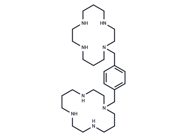
Plerixafor (AMD-3329), a chemokine receptor antagonist, blocks the binding of stromal cell-derived factor (SDF-1alpha) to the cellular receptor CXCR4.

| Pack Size | Price | Availability | Quantity |
|---|---|---|---|
| 5 mg | $42 | In Stock | |
| 10 mg | $63 | In Stock | |
| 25 mg | $113 | In Stock | |
| 50 mg | $179 | In Stock | |
| 100 mg | $323 | In Stock | |
| 200 mg | $513 | In Stock | |
| 500 mg | $818 | In Stock |
| Description | Plerixafor (AMD-3329), a chemokine receptor antagonist, blocks the binding of stromal cell-derived factor (SDF-1alpha) to the cellular receptor CXCR4. |
| Targets&IC50 | CXCL12:5.7 nM, CXCR4:44 nM |
| In vitro | Plerixafor inhibits CXCL12-mediated chemotaxis with a potency lightly better than its affinity for CXCR4. [1] Plerixafor also antagonizes SDF-1/CXCL12 ligand binding with an IC50 of 651 nM. Plerixafor inhibits SDF-1 mediated GTP-binding, SDF-1 mediated calcium flux and SDF-1 stimulated chemotaxis with IC50 of 27 nM, 572 nM and 51 nM, respectively. Plerixafor does not inhibit calcium flux against cells expressing CXCR3, CCR1, CCR2b, CCR4, CCR5 or CCR7 when stimulated with their cognate ligands, nor does Plerixafor inhibit receptor binding of LTB4. Plerixafor does not, on its own, induce a calcium flux in the CCRF–CEM cells, which express multiple GPCRs including CXCR4, CCR4 and CCR7. [2] |
| In vivo | A single topical application of Plerixafor promotes wound healing in diabetic mice by increasing cytokine production, mobilizing bone marrow EPCs, and enhancing the activity of fibroblasts and monocytes/macrophages, thereby increasing both angiogenesis and vasculogenesis. [3] Cohorts of mice are administered with PBS, IGF1, PDGF, SCF, or VEGF for five consecutive days and Plerixafor on the 5th day. The number and size of the colonies are highest in IGF1 plus Plerixafor injected mice compared to PDGF, SCF and VEGF treated groups, in combination with Plerixafor. [4] |
| Kinase Assay | In vitro biochemical assays against histone acetylases: GSK503 is profiled to assess inhibition against a panel of histone acetylases. GSK503 is dissolved in DMSO and tested in 10-dose IC50 mode with 3-fold serial dilution starting at 100 μM, with a final DMSO concentration of 2%. Anacardic Acid is used as positive control for CBP, GCN5, and pCAF and tested in 10-dose IC50 mode with 3-fold serial dilution starting at 100 μM. Curcumin is used as positive control for KAT5, MYST2/KAT7, MYST4/KAT6B, and p300, and tested in 10-dose IC50 mode with 3-fold serial dilution starting at 100 μM. Reactions are carried out at 3.08 μM Acetyl-CoA. For CBP, GCN5, MYST2/KAT7, pCAF, and p300, the substrate used is histone H3. For KAT5 and MYST4/KAT6B the substrates used are histone H2A and histone H4, respectively. |
| Cell Research | Plerixafor is dissolved in DMSO and then diluted with appropriate medium[2]. U87 mg cells are seeded in 96-well plates at the density of 6×10³ cells in 200 μL/well and treated with CXCL12, Plerixafor or with peptide R, as described in the previous "Treatments" section. MTT (5 μg/mL) is added at each time point (24, 48, 72 h) during the final 2 h of treatment. After removing cell medium, 100 μL DMSO are added and optical densities measured at 595 nm with a LT-4000MS Microplate Reader. Measurements are made in triplicates from three independent experiments[2]. |
| Alias | JM3100, AMD-3329, AMD 3100 |
| Molecular Weight | 502.78 |
| Formula | C28H54N8 |
| Cas No. | 110078-46-1 |
| Smiles | C(N1CCCNCCNCCCNCC1)c1ccc(CN2CCCNCCNCCCNCC2)cc1 |
| Relative Density. | 0.962 |
| Storage | Powder: -20°C for 3 years | In solvent: -80°C for 1 year | Shipping with blue ice. | |||||||||||||||||||||||||||||||||||
| Solubility Information | PBS: 1 mg/mL (1.99 mM), In vivo: Please add co-solvents sequentially, clarifying the solution as much as possible before adding the next one. Dissolve by heating and/or sonication if necessary. Working solution is recommended to be prepared and used immediately. Ethanol: 50 mg/mL (99.45 mM), Sonication is recommended. DMSO: < 1 mg/mL (insoluble or slightly soluble), Sonication is recommended. H2O: Insoluble | |||||||||||||||||||||||||||||||||||
Solution Preparation Table | ||||||||||||||||||||||||||||||||||||
PBS/Ethanol
Ethanol
| ||||||||||||||||||||||||||||||||||||

Copyright © 2015-2025 TargetMol Chemicals Inc. All Rights Reserved.