
Shopping Cart
- Remove All
 Your shopping cart is currently empty

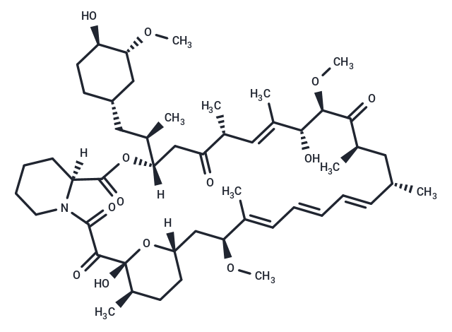
Rapamycin (AY 22989) is a natural product of macrolides, an mTOR inhibitor with specificity (HEK293 cells: IC50=0.1 nM). Rapamycin has immunosuppressive activity and induces autophagy.
| Pack Size | Price | Availability | Quantity |
|---|---|---|---|
| 10 mg | $30 | In Stock | |
| 25 mg | $40 | In Stock | |
| 50 mg | $48 | In Stock | |
| 100 mg | $63 | In Stock | |
| 200 mg | $108 | In Stock | |
| 500 mg | $198 | In Stock | |
| 1 g | $297 | In Stock | |
| 1 mL x 10 mM (in DMSO) | $43 | In Stock |
| Description | Rapamycin (AY 22989) is a natural product of macrolides, an mTOR inhibitor with specificity (HEK293 cells: IC50=0.1 nM). Rapamycin has immunosuppressive activity and induces autophagy. |
| Targets&IC50 | mTOR:0.1 nM (HEK293 cells) |
| In vitro | METHODS: Normal human renal epithelial cells HRECs were treated with Rapamycin (0.01-1000 nmol/L) for 6 days, and cell growth inhibition was detected using MTT.
RESULTS: Rapamycin dose-dependently inhibited the growth of HRECs, with a 40% reduction in cell viability at a concentration of 10 nmol/L. [1] METHODS: Human cervical cancer cells HeLa and human prostate cancer cells PC3 were treated with Rapamycin (100 nM) for 0.5-24 h, and the expression levels of target proteins were detected by Immunoprecipitation. RESULTS: Rapamycin had little effect on the expression levels of mTOR, raptor and rictor. Rapamycin significantly reduced raptor binding to mTOR at 0.5 h and rictor binding to mTOR at 24 h. Long-term treatment of cells with Rapamycin inhibited mTORC2 assembly. [2] METHODS: Human vascular endothelial cells were treated with Rapamycin (1-100 ng/mL) for 48 h, and cell migration was examined using the Wound-healing method. RESULTS: Rapamycin dose-dependently inhibited the migration of human vascular endothelial cells. [3] |
| In vivo | METHODS: To study the effect of Rapamycin on life expectancy, Rapamycin (8 mg/kg in DMSO+5% PEG-400+5% Tween-80) was administered intraperitoneally to 20-21 month old C57BL/6J mice once daily for three months.
RESULTS: Three months of Rapamycin treatment was sufficient to increase the life expectancy of middle-aged mice by 60% and improve their healthy lifespan. [4] METHODS: To determine the appropriate dose of Rapamycin for the treatment of epilepsy, Rapamycin (0.1-3 mg/kg in 4% ethanol + 5% Tween 80 + 5% PEG 400) was injected intraperitoneally into Sprague-Dawley rats once a day for four weeks. RESULTS: Only 1.0 mg/kg and 3.0 mg/kg Rapamycin inhibited p-S6. Rats treated with 0.1 and 0.3 mg/kg Rapamycin had no significant adverse effects, whereas rats treated with 1.0 and 3.0 mg/kg Rapamycin showed significant reductions in body, spleen, and thymus weights, and exhibited cognitive impairment and anxiety. The Rapamycin dose could not inhibit mTOR in the treatment of epilepsy without causing any side effects, but 1 mg/kg may be the optimal dose to inhibit mTOR in young rats with relatively few side effects. [5] |
| Kinase Assay | HEK293 cells were plated at 2-2.5 × 10^5 cells per well of a 12-well plate and serum-starved for 24 h in DMEM only. Cells were mock-treated or treated with rapamycin (0.05-50 nM), iRap (0.5-500 nM), or AP21967 (0.5-500 nM) for 15 minutes at 37 °C. Serum was added to a final concentration of 20% for 30 minutes at 37 °C. Cells were lysed as described and cell lysates were separated by SDS-PAGE. Resolved proteins were transferred to a PVDF membrane and immunoblotted with a phosphospecific primary antibody against Thr389 of p70 S6 kinase. Data were analyzed using ImageQuant and KaleidaGraph [1]. |
| Cell Research | To determine the effects of rapamycin and rapamycin plus LY294002 or UCN-01 on tumor cells, we determined cell viability after the treatments. We used a trypan blue dye exclusion assay as described previously. Tumor cells in exponential growth were harvested and seeded at 5 × 10^3 cells per well (0.1 mL) in 96-well flat-bottomed plates and incubated overnight at 37°C. The cells were then incubated for 72 hours with or without rapamycin or with rapamycin plus LY294002 or UCN-01. After the cells were collected by trypsinization, they were stained with trypan blue, and the viable cells in each well were counted. The viability of the untreated cells (the control) was considered 100%. Survival fractions were calculated from the mean cell viability of the treated cells [3]. |
| Animal Research | Animals were randomized to treatment or vehicle groups so that the mean starting body weights of each group were equal. Drug treatment began on the day of surgery or on the first day of reloading after the 14-day suspension. Rapamycin was delivered once daily by intraperitoneal injection at a dose of 1.5 mg/kg, dissolved in 2% carboxymethylcellulose. CsA was delivered once daily by subcutaneous injection at a dose of 15 mg/kg, dissolved in 10% methanol and olive oil. FK506 was delivered once daily via subcutaneous injection at a dose of 3 mg kg?1, dissolved in 10% ethanol, 10% cremophor and saline [4]. |
| Alias | Sirolimus, NSC-2260804, AY 22989 |
| Molecular Weight | 914.17 |
| Formula | C51H79NO13 |
| Cas No. | 53123-88-9 |
| Relative Density. | 1.182 g/cm3 |
| Storage | keep away from moisture,store at low temperature | Powder: -20°C for 3 years | In solvent: -80°C for 1 year | Shipping with blue ice. | ||||||||||||||||||||
| Solubility Information | Ethanol: 100 mg/mL (109 mM), Sonication is recommended. H2O: Insoluble 10% DMSO+40% PEG300+5% Tween 80+45% Saline: 10 mg/mL (10.94 mM), In vivo: Please add the solvents sequentially, clarifying the solution as much as possible before adding the next one. Dissolve by heating and/or sonication if necessary. Working solution is recommended to be prepared and used immediately. DMSO: 100 mg/mL (109 mM), Sonication is recommended. | ||||||||||||||||||||
Solution Preparation Table | |||||||||||||||||||||
Ethanol/DMSO
| |||||||||||||||||||||
 For example, your dosage is 10 mg/kg Each animal weighs 20 g, and the dosage volume is 100 μL . A total of 10 animals were administered, and the formula you used is 5%
  DMSO+30% PEG300+5% Tween 80+60% ddH2O. So your working solution concentration is 2 mg/mL。 (mother liquor concentration of 40 mg/mL), if you need to configure a concentration that exceeds the solubility of the product, please contact us first. main solution, add 300 μLPEG300 mix well and clarify, then add 50 more μL Tween 80, mix well and clarify, then add 600 more μLddH2O mix well and clarify
 Hello! How can I help you today?
Copyright © 2015-2025 TargetMol Chemicals Inc. All Rights Reserved.