Shopping Cart
- Remove All
 Your shopping cart is currently empty

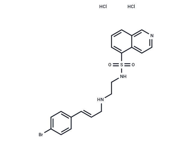
H-89 dihydrochloride (5-Isoquinolinesulfonamide) is a potent inhibitor of protein kinase A (PKA; IC50: 0.14 μM, Ki: 48 nM).

| Pack Size | Price | Availability | Quantity |
|---|---|---|---|
| 5 mg | $33 | In Stock | |
| 10 mg | $53 | In Stock | |
| 25 mg | $108 | In Stock | |
| 50 mg | $213 | In Stock | |
| 100 mg | $363 | In Stock | |
| 200 mg | $538 | In Stock | |
| 500 mg | $857 | In Stock | |
| 1 mL x 10 mM (in DMSO) | $48 | In Stock |
| Description | H-89 dihydrochloride (5-Isoquinolinesulfonamide) is a potent inhibitor of protein kinase A (PKA; IC50: 0.14 μM, Ki: 48 nM). |
| Targets&IC50 | S6K1:80 nM (cell free), PKA:48 nM (Ki, cell free) |
| In vitro | H-89 was shown to have a potent and selective inhibitory action against protein kinase A, with Ki of 0.048 microM. Pretreatment with H-89 led to a dose-dependent inhibition of the forskolin-induced protein phosphorylation, with no decrease in intracellular cyclic AMP levels in PC12D cells, and the NGF-induced protein phosphorylation was not inhibited. H-89 also significantly inhibited the forskolin-induced neurite outgrowth from PC12D cells. This inhibition also occurred when H-89 was added before the addition of dibutyryl cAMP. Pretreatment of PC12D cells with H-89 (30 microM) inhibited significantly cAMP-dependent histone IIb phosphorylation activity in cell lysates but did not affect other protein phosphorylation activity [1]. H-89 also inhibits S6K1, MSK1, ROCK-II, PKBα, and MAPKAP-K1b with IC50 values of 0.08, 0.12, 0.27, 2.6, and 2.8 μM, respectively [2]. In skinned EDL fibers of the rat, force responses to depolarization (by ion substitution) were inhibited only slightly by 10 microM H-89, a concentration more than sufficient to fully inhibit PKA. At 1-2 microM, H-89 significantly slowed the repriming rate in rat skinned fibers. With 100 microM H-89, the force response to depolarization by ion substitution was completely abolished. In intact single fibers of the flexor digitorum longus (FDB) muscle of the mouse, 1-3 microM H-89 had no noticeable effect on action-potential-mediated Ca2+ transients [3]. |
| In vivo | Different doses of H-89 (0.05, 0.1, 0.2 mg/100g) were administered intraperitoneally (i.p.), 30 min before intravenous (i.v.) infusion of PTZ (0.5% w/v). Intraperitoneal administration of H-89 (0.2 mg/100g) significantly increased seizure latency and threshold in PTZ-treated animals. Pretreatment of animals with PTX (50 and 100 mg/kg) attenuated the anticonvulsant effect of H-89 (0.2 mg/100g) in PTZ-exposed animals. H-89 (0.05, 0.2 mg/100g) prevented the epileptogenic activity of bucladesine (300 nM) with a significant increase of seizure latency and seizure threshold [4]. Treatment with H89 (10 mg/kg, administered i.p.) significantly inhibited AHR in OVA-sensitized/challenged mice, whereas it had no effect on airway responses in control mice. Treatment with H89 decreased eosinophil numbers by 80%, neutrophil numbers by 64% and lymphocyte numbers by 74% without any effect on macrophage. In the moderate model, the cell infiltrate consisted of 39.3% eosinophils, 58.5% macrophages, 1.9% neutrophils, and 0.3% lymphocytes and was entirely inhibited by H89 [5]. |
| Kinase Assay | All protein kinase activities were linear with respect to time in every incubation. Assays were performed either manually for 10 min at 30 °C in 50 μl incubations using [γ-32P]ATP or with a Biomek 2000 Laboratory Automation Workstation in a 96-well format for 40 min at ambient temperature in 25 μl incubations using [γ-33P]ATP. The concentrations of ATP and magnesium acetate were 0.1 mM and 10 mM respectively unless stated otherwise. This concentration of ATP is 5–10-fold higher than the Km for ATP of most of the protein kinases studied in the present paper, but lower than the normal intracellular concentration, which is in the millimolar range. All assays were initiated with MgATP. Manual assays were terminated by spotting aliquots of each incubation on to phosphocellulose paper, followed by immersion in 50 mM phosphoric acid. Robotic assays were terminated by the addition of 5 μl of 0.5 M phosphoric acid before spotting aliquots on to P30 filter mats. All papers were then washed four times in 50 mM phosphoric acid to remove ATP, once in acetone (manual incubations) or methanol (robotic incubations), and then dried and counted for radioactivity [2]. |
| Cell Research | After 48 h in culture, PCl2D cells are cultured in a test medium containing 30 μM H-89 for 1 h and then exposed to a fresh medium that contained both 10 μM forskolin and 30 μM H-89. Cells are scraped off with a rubber policeman and sonicated in the presence of 0.5 mL of 6% trichloroacetic acid. To extract trichloroacetic acid, 2 mL of petroleum ether is added, the preparation mixed and centrifuged at 3000 rpm for 10 min. After aspiration of the upper layer, the residue sample solution is used for determination [1]. |
| Animal Research | H89 (N-[2-(p-Bromocinnamylamino)ethyl]-5-isoquinolinesulfonamide], di-HCl Salt) (10 mg/kg) suspended in 5% DMSO in saline was administered i.p. two hours before each OVA challenge (or two hours before the last OVA challenge). Control animals received equivalent volumes (200 μl) of 5% DMSO in saline [5]. |
| Alias | Protein kinase inhibitor H-89 dihydrochloride, H 89 2HCl |
| Molecular Weight | 519.28 |
| Formula | C20H20BrN3O2S·2HCl |
| Cas No. | 130964-39-5 |
| Smiles | Cl.Cl.Brc1ccc(\C=C\CNCCNS(=O)(=O)c2cccc3cnccc23)cc1 |
| Relative Density. | no data available |
| Storage | Powder: -20°C for 3 years | In solvent: -80°C for 1 year | Shipping with blue ice. | ||||||||||||||||||||||||||||||||||||||||
| Solubility Information | H2O: < 1 mg/mL (insoluble or slightly soluble) DMSO: 60 mg/mL (115.54 mM), Sonication is recommended. 5% DMSO+95% Saline: 3.16 mg/mL (6.09 mM), In vivo: Please add the solvents sequentially, clarifying the solution as much as possible before adding the next one. Dissolve by heating and/or sonication if necessary. Working solution is recommended to be prepared and used immediately. | ||||||||||||||||||||||||||||||||||||||||
Solution Preparation Table | |||||||||||||||||||||||||||||||||||||||||
5% DMSO+95% Saline/DMSO
DMSO
| |||||||||||||||||||||||||||||||||||||||||

Copyright © 2015-2025 TargetMol Chemicals Inc. All Rights Reserved.