
Shopping Cart
- Remove All
 Your shopping cart is currently empty

BI-D1870 is a highly specific, blood-brain-permeable, and ATP-competitive inhibitor of the N-terminal AGC kinase domain of RSK, with IC50 values of 31 nM, 24 nM, 18 nM, and 15 nM for RSK1, RSK2, RSK3, and RSK4.
| Pack Size | Price | Availability | Quantity |
|---|---|---|---|
| 1 mg | $48 | In Stock | |
| 2 mg | $71 | In Stock | |
| 5 mg | $139 | In Stock | |
| 10 mg | $217 | In Stock | |
| 25 mg | $353 | In Stock | |
| 50 mg | $497 | In Stock | |
| 100 mg | $693 | In Stock | |
| 500 mg | $1,450 | In Stock | |
| 1 mL x 10 mM (in DMSO) | $119 | In Stock |
| Description | BI-D1870 is a highly specific, blood-brain-permeable, and ATP-competitive inhibitor of the N-terminal AGC kinase domain of RSK, with IC50 values of 31 nM, 24 nM, 18 nM, and 15 nM for RSK1, RSK2, RSK3, and RSK4. |
| Targets&IC50 | RSK3:18 nM (cell free), RSK2:24 nM (cell free), RSK1:31 nM (cell free), RSK4:15 nM (cell free) |
| In vitro | METHODS: HEK-293 cells were incubated with BI-D1870 (10 μM, 15 min, 4 h total) To examine whether BI-D1870 could inhibit RSK activity in cells, the effect of BI-D1870 on RSK-catalyzed phosphorylation of its known substrates in HEK-293 cells was investigated using PMA as an agonist to activate ERK1/ERK2 and RSK isoforms. RESULTS Incubation of cells with BI-D1870 significantly inhibited PMA-induced phosphorylation of GSK3α and GSK3β. In contrast, BI-D1870 had little effect on PMA-induced activation of ERK1/ERK2 (catalyzed by Raf and MKK1) or phosphorylation of CREB at Ser at any time point. [1] METHODS: Oral cancer cell lines (SCC2095, SCC4, SCC9, Ca922 and HSC-3) and NHOK cells were treated with BI-D1870 (1, 2, 3, 4, 5.28 or 48 hours), and the growth of these cells was detected by MTT. RESULTS BI-D1870 showed a dose-responsive antiproliferative effect on OSCC cells. [3] METHODS: Cells transfected with immunofluorescent GFP-LC3 were treated with BI-D1870 (0.5, 2 μM) for 48 hours and observed under a confocal microscope; DAPI staining was used to locate the nucleus, and Western blot of LC3B was observed in cells treated with BI-D1870 for 48 hours. RESULTS BI-D1870 induced autophagy, and Western blot of LC3B-II showed that this induction was dose-dependent. [3] |
| In vivo | METHODS: Two days after immunization with MOG peptide, BI-D1870 (0.5 mg/kg) was injected intraperitoneally into mice and repeated every other day for 11 days. RESULTS Mice showed delayed neurological deficits; BI-D1870 treatment had a moderate protective effect against weight loss. [4] |
| Kinase Assay | Purified His6–RSK1, His6–RSK2 or GST–RSK21–389:S381E (1–2 units/ml) were assayed for 10 min at 30 °C in a 50 μl assay mixture in Buffer A containing 30 μM substrate peptide (KEAKEKRQEQIAKRRRLSSLRASTSKSGGSQK), 10 mM magnesium acetate and 100 μM of [γ-32P]ATP. Reactions were terminated and analyzed as described previously. The amount of enzyme that catalyzed the phosphorylation of 1 nmol of substrate peptide in 1 min was termed one unit. In order to assay RSK and MSK1 in HEK-293 or Rat-2 cell lysates, these kinases were immunoprecipitated from the cell lysates (0.1 mg of lysate protein for RSK and 0.3 mg for MSK1) and assayed as described previously, except that for RSK assays the immunoprecipitates were washed twice with Buffer A containing 1 mM ATP and twice with Buffer A prior to the assay, as a precaution to ensure dissociation of BI-D1870 from the RSK isoforms [1]. |
| Cell Research | The rat embryo fibroblast cell line, Rat-2 cells were cultured on 10 cm-diameter dishes in Dulbecco's Modified Eagle's medium supplemented with 10% (v/v) FBS. HEK-293 cells were cultured on 10 cm-diameter dishes in Dulbecco's Modified Eagle's medium supplemented with 10% FBS and 1×antimycotic/antibiotic solution. Prior to stimulation, cells were cultured in the absence of serum for 16 h. Inhibitors were dissolved in DMSO at a 1000-fold higher concentration than they were used at. These inhibitors, or the equivalent volume of DMSO as a control, were added to the tissue culture medium 30 min prior to stimulation unless indicated otherwise. The final concentration of DMSO in the culture medium was 0.1% and had no effect on agonist-induced activation or phosphorylation of any of the substrates examined. The cells were stimulated with the indicated agonists and lysed in 1 ml of ice-cold Lysis Buffer and centrifuged at 16000 g at 4 °C for 5 min. The supernatants were frozen in liquid nitrogen and stored at ?80 °C until use. Protein concentrations were determined using the Bradford method with BSA as the standard [1]. |
| Animal Research | Myelin oligodendrocyte glycoprotein (MOG) peptide 35–55. (MEVGWYRSPFSRVVHLYRNGK) (BEX) was used to induce EAE in C57/BL6J mice. Mice were injecteds.c. with 200 g of MOG peptide in100 L of PBS emulsified in 100 L complete Freund's adjuvant (CFA) that was further supplemented with five mg mL?1 Mycobacterium tuberculosis (H37Ra). In addition, 500 ng pertussis toxin was injected i.p. on days zero and two. The RSK inhibitor (BI-D1870; 0.5 mg kg?1) was injected i.p. into mice two days after immunization with MOG peptide, and injection was repeated every other day for 11 days. Mice that received only dimethyl sulfoxide (DMSO) solution were used as controls. Paralysis was evaluated according to the following scale: zero, no disease; one, tail limpness; two, hind limb weakness; three, hind limb paralysis; four, forelimb weakness; five, quadriplegia; six, death. For histological analysis, CNS samples were fixed with 4% paraformaldehyde and sliced at 4 m, and then hematoxylin & eosin (H & E) staining was performed [4]. |
| Molecular Weight | 391.42 |
| Formula | C19H23F2N5O2 |
| Cas No. | 501437-28-1 |
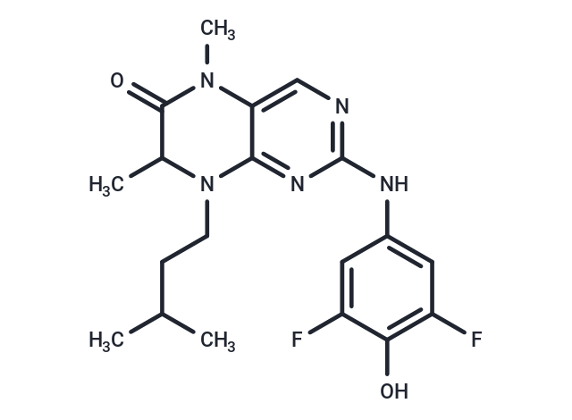
| Smiles | CC(C)CCN1C(C)C(=O)N(C)c2cnc(Nc3cc(F)c(O)c(F)c3)nc12 |
| Relative Density. | 1.297 |
| Storage | Powder: -20°C for 3 years | In solvent: -80°C for 1 year | Shipping with blue ice. |
| Solubility Information | Ethanol: < 1 mg/mL (insoluble or slightly soluble) H2O: < 1 mg/mL (insoluble or slightly soluble) DMSO: 6.88 mg/mL (17.56 mM), Sonication is recommended. 10% DMSO+40% PEG300+5% Tween 80+45% Saline: 7.2 mg/mL (18.39 mM), suspension.In vivo: Please add the solvents sequentially, clarifying the solution as much as possible before adding the next one. Dissolve by heating and/or sonication if necessary. Working solution is recommended to be prepared and used immediately. |
Solution Preparation Table | |
 For example, your dosage is 10 mg/kg Each animal weighs 20 g, and the dosage volume is 100 μL . A total of 10 animals were administered, and the formula you used is 5%
  DMSO+30% PEG300+5% Tween 80+60% ddH2O. So your working solution concentration is 2 mg/mL。 (mother liquor concentration of 40 mg/mL), if you need to configure a concentration that exceeds the solubility of the product, please contact us first. main solution, add 300 μLPEG300 mix well and clarify, then add 50 more μL Tween 80, mix well and clarify, then add 600 more μLddH2O mix well and clarify
 Hello! How can I help you today?
Copyright © 2015-2025 TargetMol Chemicals Inc. All Rights Reserved.