Shopping Cart
- Remove All
 Your shopping cart is currently empty

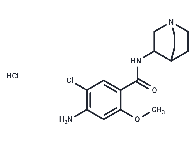
Zacopride is a highly potent 5-HT3 receptor antagonist (Kd = 0.38 nM) and 5-HT4 receptor agonist (Ki = 373 nM). It also is a selective IK1 channel agonist.

| Pack Size | Price | Availability | Quantity |
|---|---|---|---|
| 1 mg | $38 | In Stock | |
| 5 mg | $89 | In Stock | |
| 10 mg | $147 | In Stock | |
| 25 mg | $297 | In Stock | |
| 50 mg | $478 | In Stock | |
| 100 mg | $689 | In Stock | |
| 200 mg | $979 | In Stock | |
| 1 mL x 10 mM (in DMSO) | $98 | In Stock |
| Description | Zacopride is a highly potent 5-HT3 receptor antagonist (Kd = 0.38 nM) and 5-HT4 receptor agonist (Ki = 373 nM). It also is a selective IK1 channel agonist. |
| Targets&IC50 | 5-HT4:373 nM (Ki, cell free), 5-HT3:0.38 nM (Kd, cell free) |
| In vitro | Zacopride (0.1-10 μmole/L) dose-dependently enhanced the IK1 current in isolated rat cardiomyocytes, had no effects on other ion channels, transporters, or pumps [2]. In Langendorff perfused rat hearts, the antiarrhythmic effect of 1 μmol/L zacopride were reversed by 1 μmol/L BaCl2, a blocker of IK1 channel. In Kir2.x transfected Chinese hamster ovary (CHO) cells, zacopride activated the Kir2.1 homomeric channel but not the Kir2.2 or Kir2.3 channels [3]. |
| In vivo | In all the three treatment modes, zacopride (15 μg/kg) inhibited MI-induced ventricular tachyarrhythmias, as shown by significant decreases in the premature ventricular contraction (PVC) and the duration and incidence of VT or VF [3]. Zacopride enhanced exploratory behaviour and social interaction in the mouse and rat models and antagonized the defensive response in the marmoset, zacopride being 100 times more potent than diazepam [4]. |
| Kinase Assay | [3H] GR113808 binding studies were performed. Membranes were incubated at 37°C for 30 min in 0.5 ml of 50 mM Hepes pH 7.4 buffer, containing 0.1 nM [3H]GR113808, a specific ligand for 5-HT4 receptor, for the competition binding studies and varying concentrations of the radioligand for the saturation studies. Non-specific binding was defined with 30 μM 5-HT. For all binding studies, the incubation was terminated by rapid filtration and washing with ice-cold Hepes buffer through GF/B filters using a Brandel Cell Harvester. Radioactivity of the filter was counted [1]. |
| Cell Research | In brief, a rat heart was quickly removed and mounted via the aorta on an 80-cm H2O high Langendorff retrograde perfusion apparatus. The heart was perfused first with oxygenated Ca2+-free Tyrode solution (at 37°C) for approximately 7–8 minutes, then with collagenase P (0.1 g/L) Tyrode solution for about 15 minutes. The composition of the Tyrode solution was (in millimoles per liter): NaCl 135, KCl 5.4, CaCl2 1.8, MgCl2 1.0, NaH2PO4 0.33, HEPES 10, glucose 10, pH adjusted to 7.3–7.4 with NaOH. When the tissue was well digested, the left ventricle was separated, minced and filtrated in KB solution with the following composition (in millimoles per liter): KOH 85, L-glutamic acid 50, KCl 30, MgCl2 1.0, KH2PO4 30, glucose 10, taurine 20, HEPES 10, ethylene glycol tetraacetic acid (EGTA), 0.5, pH adjusted to 7.4 with KOH. The isolated myocytes were stored at room temperature (23–25°C) at least 2 hours before use. The cell suspension was then transferred to a chamber mounted on an inverted microscope and superfused with the Tyrode solution. The whole-cell recording was performed with an amplifier Axopatch 200B. When filled with the pipette solution, the electrode resistance was maintained at 2–5 MΩ except for 1–1.5 MΩ for the Na+ current recording. The current signal was filtered at 2 kHz and sampled at 10– 20 kHz. Current was recorded in the voltage clamp mode, and membrane potentials were recorded in the current clamp mode at a frequency of 1.0 Hz. The baseline currents or APs were recorded first, then current values were recorded in the presence of zacopride at different concentrations (0.1, 1.0, 10 μmole/L). Ionic currents were corrected for cell capacitance and expressed in terms of current density (picoamperes/picofarads). All the experiments were conducted at room temperature (23–25°C), except at 36°C for AP recordings. The series resistance was 5.7 ± 2.2 MΩ, and the cell capacitance and pipette series resistance were mostly compensated (usually >70%) [2]. |
| Animal Research | Aconitine was administered into the exposed vena jugularis in the course of 10 seconds at a dosage of 30 μg/kg (0.046 μmole/kg). Then, 0.2 mL normal saline (NS) or zacopride at a dosage of 25 μg/kg (dissolved in NS, ~0.2 mL) or lidocaine at 7.5 mg/kg (dissolved in NS, ~0.2 mL) were administered immediately into the exposed left femoral vein (n = 8 in each group). In the pilot experiments, 25 μg/kg of zacopride and 7.5 mg/kg of lidocaine showed the optimal antiarrhythmic effects and fewer proarrhythmic side effects. The ECG signals were recorded continuously for a total of 1 hour after the administration of aconitine, and the arrhythmias were evaluated. Various arrhythmic activities, such as DADs, triggered activity (TA), premature ventricular contraction (PVC), ventricular tachycardia (VT), and ventricular fibrillation (VF) were monitored in the observed time window. Parameters of various arrhythmias, such as the episodes and the average duration of each type of arrhythmias, the incidence rate, and the total duration of the arrhythmias among groups, were calculated [2]. |
| Molecular Weight | 346.26 |
| Formula | C15H20ClN3O2.HCl |
| Cas No. | 101303-98-4 |
| Smiles | Cl.COc1cc(N)c(Cl)cc1C(=O)NC1CN2CCC1CC2 |
| Relative Density. | no data available |
| Storage | Powder: -20°C for 3 years | In solvent: -80°C for 1 year | Shipping with blue ice. | |||||||||||||||||||||||||||||||||||
| Solubility Information | DMSO: 60 mg/mL (173.28 mM), Sonication is recommended. | |||||||||||||||||||||||||||||||||||
Solution Preparation Table | ||||||||||||||||||||||||||||||||||||
DMSO
| ||||||||||||||||||||||||||||||||||||

Copyright © 2015-2025 TargetMol Chemicals Inc. All Rights Reserved.