Shopping Cart
- Remove All
 Your shopping cart is currently empty

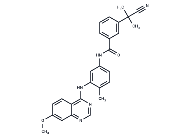
AZ304 is an ATP-competitive dual BRAF kinase inhibitor, effectively inhibiting BRAF (WT), BRAF (V600E), and wild-type CRAF [IC50s: 79/38/68 nM].

| Pack Size | Price | Availability | Quantity |
|---|---|---|---|
| 10 mg | $35 | In Stock | |
| 25 mg | $59 | In Stock | |
| 50 mg | $98 | In Stock | |
| 100 mg | $158 | In Stock | |
| 1 mL x 10 mM (in DMSO) | $37 | In Stock |
| Description | AZ304 is an ATP-competitive dual BRAF kinase inhibitor, effectively inhibiting BRAF (WT), BRAF (V600E), and wild-type CRAF [IC50s: 79/38/68 nM]. |
| Targets&IC50 | C-Raf:68 nM (cell free), B-Raf:79 nM (cell free), p38:6 nM (cell free), CSF1R:35 nM (cell free), B-Raf (V600E):38 nM (cell free) |
| In vitro | AZ304 showed potent inhibitory activities to the kinase domains of wild type BRAF, V600E mutant BRAF and wild type CRAF in vitro, with IC50 values of 79 nM, 38 nM, and 68 nM, respectively. Consistent with BRAF kinase inhibition in vitro, AZ304 potently reduced ERK phosphorylation (p-ERK), with a mean EC50 of 65 nM in the V600E mutant BRAF containing melanoma cell line A375; an EC50 of 60 nM was obtained for the wild type BRAF melanoma cell line SK-MEL-31. AZ304 markedly inhibited cell proliferation in mutant BRAF cancer cell lines and effectively reduced cell growth in selected cell lines harboring wild type BRAF/RAS or mutant RAS. The GI50 values ranged from 0.08–7.72 μM in mutant BRAF cell lines, 0.43–11.7 μM in wild type BRAF/RAS cell lines, and 0.9–16.66 μM in mutant RAS cell lines. |
| In vivo | Compared with vehicle-treated controls, treatment with AZ304 or Cetuximab alone resulted in reduced tumor growth in both xenograft models. Furthermore, the AZ304 and Cetuximab combination caused dramatic tumour growth inhibition and even shrinking in the Caco-2 xenograft model. |
| Kinase Assay | Briefly, the kinase activity of BRAF WT, BRAF V600E, or CRAF was measured using an AlphaScreen assay monitoring MEK1/2 phosphorylation at Ser217/221. For CDK2 and CDK4 kinases, activity was also measured by AlphaScreen, monitoring phosphorylation of biotin Rb peptide at Ser780. Similarly, MAP3K7, CSF1R, and JAK2 kinase activity were measured by phosphorylation of their biotinylated substrates MKK6 kinase-dead protein at Ser271/Thr275, or tyrosine phosphorylation of pEY or Tyk2 Tyr1054/1055 peptides, respectively. CSK, IGF1R, EGFR, FGFR1 and SRC kinase activity was measured using a sandwich ELISA detecting phosphorylated poly EAY peptide with a HRP conjugated phosphotyrosine antibody and TMB substrate, while p38 kinase activity was measured by monitoring phosphorylation of MBP protein with radiolabeled 33P-ATP in a filter binding format. All assays were screened under respective ATP Km conditions and inhibitor IC50s were derived from either 5 (RAF kinases, CDK2, CDK4, MAP3K7, JAK2), 10 (CSK, IGF1R, EGFR, FGFR1, SRC) or 11 (p38, CSF1R) point compound dose response. |
| Cell Research | Briefly, the cells were treated with DMSO or multiple concentrations of AZ304 for 3 days. The cell growth was determined using the CellTiter 96 Aqueous One Cell Proliferation Assay. Percentage of net growth at day 3 (100%) relative to day 0 (0%) was calculated and the concentration of compound required to inhibit growth by 50% (GI50) determined. The assays were done in triplicate across different plates. The proliferation assays in selected colorectal cancer cell lines were measured using an MTT assay. First of all, cultured cells were seeded into 96-well plates (2000–5000 cells per well). After incubation for 24 h, the cells were pretreated with DMSO or AZ304 for 1 h. Then the indicated doses of Cetuximab were added. Cells were incubated for a further 48 or 72 h. For EGF stimulating assay, cells were incubated in reduced serum medium overnight and then treated with AZ304 or AZ304 + EGF (20 ng/ml) for 72 h. Twenty microliters of MTT solution (5 mg/ml) was added to each well followed by 4 h incubation at 37 °C. The cell culture medium was removed and the cells were lysed in 200 μl DMSO and the results were measured using a microplate reader. |
| Animal Research | Female 4–6 weeks old athymic BALB/c nude mice were purchased from Shanghai SLAC Laboratory Animal Centre. RKO/Caco-2 cells (1 × 10^7) in 200 μl PBS were injected subcutaneously into the right scapular region of mice. After the average tumour size reached 150 – 200 mm^3, animals were randomly divided into 4 groups, each containing three mice and were treated with vehicle only (CON) which orally received 0.5% HPMC and injected with 0.9% saline, AZ304 only (AZ304 dissolved in 0.5% HPMC, 10 mg/kg by oral gavage twice daily), Cetuximab only (40 mg/kg by intraperitoneal injection twice per week), or their combination (A+C) for 10 days. Tumors were measured with a caliper every 2 days, so did body weights. Tumor volume was calculated using the formula V = 1/2 (width^2 × length). Mice were terminated by CO2 inhalation when the tumor diameters reached 1.5 cm, according to the protocol filed with the Guidance of Institutional Animal Care and Use Committee of China Medical University. |
| Molecular Weight | 451.52 |
| Formula | C27H25N5O2 |
| Cas No. | 942507-42-8 |
| Smiles | COc1ccc2c(Nc3cc(NC(=O)c4cccc(c4)C(C)(C)C#N)ccc3C)ncnc2c1 |
| Relative Density. | 1.270 g/cm3 (Predicted) |
| Storage | Powder: -20°C for 3 years | In solvent: -80°C for 1 year | Shipping with blue ice. | ||||||||||||||||||||||||||||||||||||||||
| Solubility Information | DMSO: 50 mg/mL (110.74 mM), Sonication is recommended. H2O: Insoluble Ethanol: 4 mg/mL (8.86 mM), Sonication is recommended. | ||||||||||||||||||||||||||||||||||||||||
Solution Preparation Table | |||||||||||||||||||||||||||||||||||||||||
Ethanol/DMSO
DMSO
| |||||||||||||||||||||||||||||||||||||||||

Copyright © 2015-2025 TargetMol Chemicals Inc. All Rights Reserved.