Shopping Cart
- Remove All
 Your shopping cart is currently empty

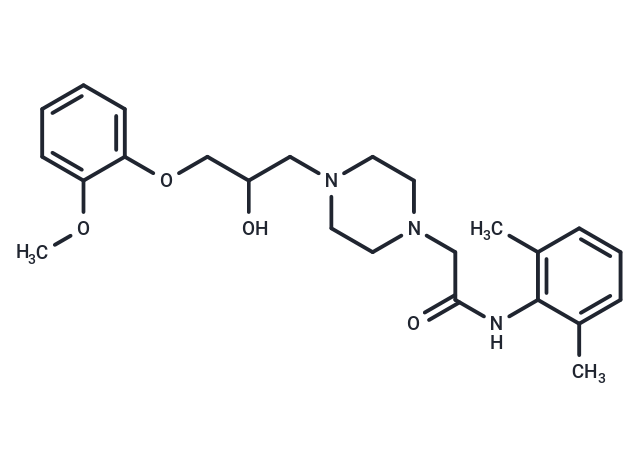
Ranolazine (RS 43285-003) is a calcium uptake inhibitor via the sodium/calcium channel, used to treat chronic angina. It affects the sodium-dependent calcium channels during myocardial ischemia in rabbits by altering the intracellular sodium level.

| Pack Size | Price | Availability | Quantity |
|---|---|---|---|
| 50 mg | $57 | In Stock | |
| 100 mg | $81 | In Stock | |
| 200 mg | $125 | In Stock | |
| 500 mg | $212 | In Stock | |
| 1 mL x 10 mM (in DMSO) | $50 | In Stock |
| Description | Ranolazine (RS 43285-003) is a calcium uptake inhibitor via the sodium/calcium channel, used to treat chronic angina. It affects the sodium-dependent calcium channels during myocardial ischemia in rabbits by altering the intracellular sodium level. |
| Targets&IC50 | IKr:12 μM (IC50), INa:6 μM (IC50) |
| In vitro | In the absence and presence of IK-blocking drugs, when late INa is increased, Ranolazine inhibits the late component of INa and attenuates prolongation of action potential duration. Ranolazine (10 mM) reduces by 89% of the 13.6-fold increase in variability of APD caused by 10 nM ATX-II. Ranolazine is found to bind more tightly to the inactivated state than the resting state of the sodium channel underlying I(NaL), with apparent dissociation constants K(dr)=7.47 mM and K(di)=1.71 mM, respectively. Ranolazine(5 mM and 10 mM) reversibly shortens the duration of TCs and abolishes the after contraction. |
| In vivo | In dog left ventricular myocytes, Ranolazine significantly and reversibly stimulate the action potential duration (APD) of shortened myocytes at 0.5 or 0.25 Hz in a concentration-dependent manner. In rat hearts, Ranolazine (10 mM) significantly increased 1.5-fold to 3-fold under glucose oxidation conditions. Ranolazine (10 mM) also increased glucose oxidation (high calcium, low FA; 15 ml/min) in Langendorff hearts in normoxic rats. Ranolazine significantly improves the function of the reperfused ischemic working heart, which is associated with a significant increase in glucose oxidation function. |
| Kinase Assay | In vitro kinase assay for CDK1 and Aurora kinases:CDK1 kinase activity is tested by the CDK1/cyclin B complex purified from baculovirus to phosphorylate a biotinylated peptide substrate containing the consensus phosphorylation site for histone H1, which is phosphorylated in vivo by CDK1. Inhibition of CDK1 activity is measured by observing a decreased amount of 33P-γ-ATP incorporation into the immobilized substrate in streptavidin-coated 96-well scintillating microplates. CDK1 enzyme is diluted in 50 mM Tris-HCl (pH 8), 10 mM MgCl2, 0.1 mM Na3VO4, 1 mM DTT, 1% DMSO, 0.25 μM peptide, 0.1 μCi per well 33P-γ-ATP, and 5 μM ATP in the presence or absence of various concentrations of JNJ-7706621 and incubated at 30 °C for 1 hour. The reaction is terminated by washing with PBS containing 100 mM EDTA and plates are counted in a scintillation counter. IC50 is determined by Linear regression analysis of the percent inhibition by JNJ-7706621.The Aurora kinase activity is measured with 10 μM ATP and a peptide containing a dual repeat of the kemptide phosphorylation motif. |
| Alias | RS 43285-003, Ranexa, CVT 303 |
| Molecular Weight | 427.54 |
| Formula | C24H33N3O4 |
| Cas No. | 95635-55-5 |
| Smiles | COC1=CC=CC=C1OCC(O)CN1CCN(CC(=O)NC2=C(C)C=CC=C2C)CC1 |
| Relative Density. | 1.174 g/cm3 |
| Storage | Powder: -20°C for 3 years | In solvent: -80°C for 1 year | Shipping with blue ice. | ||||||||||||||||||||||||||||||||||||||||
| Solubility Information | H2O: < 1 mg/mL (insoluble or slightly soluble) Ethanol: 16 mg/mL (37.42 mM), Sonication is recommended. DMSO: 60 mg/mL (140.34 mM), Sonication is recommended. | ||||||||||||||||||||||||||||||||||||||||
Solution Preparation Table | |||||||||||||||||||||||||||||||||||||||||
Ethanol/DMSO
DMSO
| |||||||||||||||||||||||||||||||||||||||||

Copyright © 2015-2025 TargetMol Chemicals Inc. All Rights Reserved.