Shopping Cart
- Remove All
 Your shopping cart is currently empty

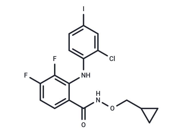
CI-1040 (PD 184352) (PD184352) is an ATP non-competitive MEK1/2 inhibitor (IC50: 17 nM).

| Pack Size | Price | Availability | Quantity |
|---|---|---|---|
| 5 mg | $48 | In Stock | |
| 10 mg | $77 | In Stock | |
| 25 mg | $150 | In Stock | |
| 50 mg | $231 | In Stock | |
| 100 mg | $385 | In Stock | |
| 200 mg | $482 | In Stock | |
| 1 mL x 10 mM (in DMSO) | $53 | In Stock |
| Description | CI-1040 (PD 184352) (PD184352) is an ATP non-competitive MEK1/2 inhibitor (IC50: 17 nM). |
| Targets&IC50 | MEK1:17 nM (cell free), MEK2:17 nM (cell free) |
| In vitro | PD 184352 (CI-1040) also substantially reduced steady-state levels of phosphorylated MAPK (pMAPK) in a diverse panel of tumor lines grown in the presence of serum. Treatment of colon 26 cells for 1 hour with 1 μM PD 184352 produced a reduction of pMAPK levels of more than 75%. Treatment of colon 26 cells with 10 μM PD 184352 did not inhibit the phosphorylation of Jun kinase, p38 kinase or AKT [1]. The IC50 for inhibition of MKK1 by PD 184352 was 0.3 μM, 15-fold higher than the concentration required to inhibit the EGF-induced activation of ERK2 in Swiss 3T3 cells. The activation of MKK1 in cells was inhibited by 50% at 2 nM PD 184352 [2]. CI-1040 induced apoptosis and inhibited proliferation in U-937 cells in a dose and time-dependent manner. CI-1040 induced a significant increase in PUMA mRNA and protein levels. Knockdown of PUMA by PUMA siRNA transfection inhibited CI-1040-induced apoptosis and proliferation inhibition in U-937 cells [3]. |
| In vivo | The tumors were excised at 1 hour and 6 hours after treatment with PD 184352 (150 mg/kg, i.p. or p.o.). Treatment with PD 184352 completely suppressed MAPK phosphorylation by either route of administration for at least 6 hours. MAPK phosphorylation returned at 12 hours after dosing and attained control levels by 24 hours [1]. In vivo, the systemic administration of the MEK inhibitor CI-1040 reduced adenoma formation to a third and significantly restored lung structure. The proliferation rate of lung cells of mice treated with CL-1040 was decreased without any obvious effects on the differentiation of pneumocytes [4]. |
| Kinase Assay | All protein kinase activities were linear with respect to time in every incubation. Assays were performed either manually for 10 min at 30 °C in 50 μl incubations using [γ-32P]ATP, or with a Biomek 2000 Laboratory Automation Workstation in a 96-well format for 40 min at ambient temperature in 25 μl incubations using [γ-33P]ATP. The concentrations of ATP and magnesium acetate were 0.1 mM and 10 mM respectively, unless stated otherwise. This concentration of ATP is 5–10-fold higher than the Km for ATP of most of the protein kinases studied in the present paper, but lower than the normal intracellular concentration, which is in the millimolar range. All assays were initiated with MgATP. Manual assays were terminated by spotting aliquots of each incubation on to phosphocellulose paper, followed by immersion in 50 mM phosphoric acid. Robotic assays were terminated by the addition of 5 μl of 0.5 M phosphoric acid before spotting aliquots on to P30 filter mats. All papers were then washed four times in 50 mM phosphoric acid to remove ATP, once in acetone (manual incubations) or methanol (robotic incubations), and then dried and counted for radioactivity [2]. |
| Cell Research | Cells were planted seeded in T-75 cm2 flasks and treated the next day for 24 h with either DMSO or PD 184352. Single-cell suspensions were collected, and pellets were fixed in ice-cold ethanol (70%) for 30 min. After centrifugation of the samples, propidium iodide (50 μg/ml) and RNase (30 units/ml) were added to the pellets for 20 min at 37 °C. After filtration, samples were analyzed by flow cytometry [1]. |
| Animal Research | Tumor fragments (approximately 3 mm^3 in size) were implanted subcutaneously into the right axillae of CD2F1 male mice (colon 26 studies) or female nude mice (HT-29 studies) 4–6 weeks old. Treatment was administered by gavage or intraperitoneally and was initiated either the day after tumor implantation (colon 26) or when tumors reached approximately 200 mg in size (HT-29). PD 184352 was prepared in a vehicle of 10% Cremophore EL, 10% ethanol and 80% water. Tumor size was evaluated periodically by caliper measurements, generally three times per week. Percent tumor growth inhibition was calculated as [(T–C)/number of days of treatment] × 100, with T and C being defined as the time required for treated and control tumors, respectively, to reach 750 mg (colon 26) or to reach twofold growth (HT-29) [1]. |
| Alias | PD 184352 |
| Molecular Weight | 478.66 |
| Formula | C17H14ClF2IN2O2 |
| Cas No. | 212631-79-3 |
| Smiles | Fc1ccc(C(=O)NOCC2CC2)c(Nc2ccc(I)cc2Cl)c1F |
| Relative Density. | 1.747 g/cm3 (Predicted) |
| Storage | Powder: -20°C for 3 years | In solvent: -80°C for 1 year | Shipping with blue ice. | ||||||||||||||||||||||||||||||||||||||||
| Solubility Information | Ethanol: 12 mg/mL (25.07 mM), Sonication is recommended. DMSO: 47.9 mg/mL (100.07 mM), Sonication is recommended. | ||||||||||||||||||||||||||||||||||||||||
Solution Preparation Table | |||||||||||||||||||||||||||||||||||||||||
Ethanol/DMSO
DMSO
| |||||||||||||||||||||||||||||||||||||||||

Copyright © 2015-2025 TargetMol Chemicals Inc. All Rights Reserved.