Shopping Cart
- Remove All
 Your shopping cart is currently empty

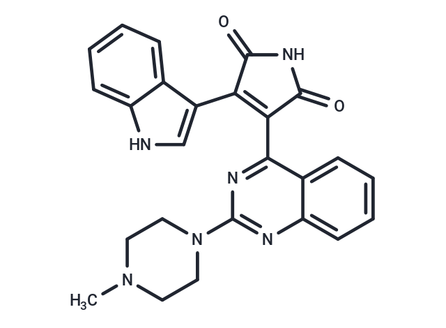
Sotrastaurin (AEB071) is a potent pan-PKC inhibitor with Kis of 0.95 nM for PKCα, 0.64 nM for PKCβI, 2.1 nM for PKCδ, 3.2 nM for PKCε, 1.8 nM for PKCη, and 0.22 nM for PKCθ.

| Pack Size | Price | Availability | Quantity |
|---|---|---|---|
| 1 mg | $45 | In Stock | |
| 5 mg | $97 | In Stock | |
| 10 mg | $169 | In Stock | |
| 25 mg | $329 | In Stock | |
| 50 mg | $583 | In Stock | |
| 100 mg | $832 | In Stock | |
| 500 mg | $1,660 | In Stock | |
| 1 mL x 10 mM (in DMSO) | $117 | In Stock |
| Description | Sotrastaurin (AEB071) is a potent pan-PKC inhibitor with Kis of 0.95 nM for PKCα, 0.64 nM for PKCβI, 2.1 nM for PKCδ, 3.2 nM for PKCε, 1.8 nM for PKCη, and 0.22 nM for PKCθ. |
| Targets&IC50 | PKCα:0.95 nM(Ki, cell free), PKCη:1.8 nM(Ki, cell free), PKCδ:2.1 nM(Ki, cell free), PKCθ:0.22 nM(Ki, cell free), PKCβ1:0.64 nM(Ki, cell free), PKCε:3.2 nM(Ki, cell free) |
| In vitro | In cell-free kinase assays, Sotrastaurin (AEB071) inhibited PKC, with K(i) values in the subnanomolar to low nanomolar range. Upon T-cell stimulation, AEB071 markedly inhibited in situ PKC catalytic activity. In primary human and mouse T cells, AEB071 treatment effectively abrogated at low nanomolar concentration markers of early T-cell activation [1]. Growth inhibition was observed in GNAQ/GNA11-mutant cells with AEB071 versus no activity in wild-type cells. In the GNAQ-mutant cells, AEB071 decreased phosphorylation of myristoylated alanine-rich C-kinase substrate, a substrate of PKC, along with ERK1/2 and ribosomal S6, but persistent AKT activation was present [2]. |
| In vivo | Daily oral dosing of Sotrastaurin (80 mg/kg, tid) resulted in statistically significant inhibition of tumor growth compared with vehicle-treated animals, corresponding to 17% tumor volume change, treated over the control group [2]. The combination therapy resulted in a significantly enhanced reduction in tumor volume when compared to either AEB071 or BYL719 alone. There was even a greater effect when compared to vehicle control [3]. |
| Kinase Assay | Classical and novel PKC isotypes were assayed by scintillation proximity assay technology. In brief, the assay was performed in 20 mM Tris-HCl buffer, pH 7.4, and 0.1% bovine serum albumin by incubating 1.5 μM of the peptide substrate with 10 μM [33P]ATP, 10 mM Mg(NO3)2, 0.2 mM CaCl2, and PKC at a protein concentration varying from 25 to 400 ng/ml, and lipid vesicles containing 30 mol% phosphatidylserine, 5 mol% diacylglycerol (DAG), and 65 mol% phosphatidylcholine at a final lipid concentration of 0.5 μM. Incubation was performed for 60 min at room temperature. The reaction was stopped by adding 50 μl of a mixture containing 100 mM EDTA, 200 μM ATP, 0.1% Triton X-100, and 0.375 μg/well streptavidin-coated scintillation proximity assay beads in PBS without Ca2+ and Mg2+. Incorporated radioactivity was measured in a MicroBetaTrilux counter for 1 min. In situ Thr-219 autophosphorylation status analysis of PKC was done by a phospho-site-specific antibody [1]. |
| Cell Research | Jurkat cells (5×10^6 cells) were pretreated for 4 h with 500 nM AEB071 and loaded for 30 min at 37°C in the dark with 5 μM fura-2 acetoxymethyl ester. Dye excess was removed by washing in Hanks' balanced salt solution. Samples were prewarmed to 37°C and baseline Ca2+ levels were determined for 100 s on a Spex Fluorolog 2 spectrofluorometer equipped with two excitation monochrometers and a Cooper system. At this point, anti-CD3 antibody was added to a final concentration of 10 μg/ml, and data were collected over 6.5 min. The maximal and minimal Ca2 levels were determined by adding an excess of ionomycin and EGTA. Experiments were performed at least four times with similar outcomes [1]. |
| Animal Research | 6–8 week nu/nu SCID female mice bearing subcutaneously injected 92.1 tumors (7 mice/group) of 100mm3 diameter were treated with vehicle, AEB071 (80mg/kg/d) TID and or BYL719 orally (50mg/kg/d) QD as single agents and in combination, 5 days/week for 2 weeks. After 2 weeks, two animals from each group were sacrificed and tumors were collected to analyze for Western blot. For Omm1 xenografts, 6–8 weeks athymic female mice bearing subcutaneously injected Omm1 tumors (7 mice/group) of 100 mm3 diameter were treated with vehicle, AEB071 (80mg/kg/d) TID and or BYL719 orally (50mg/kg/d) QD as single agents and in combination, 5 days/week for 3 weeks. Tumors were homogenized with grinding resins kits as per manufacturer's instructions. Tumors were collected to analyze for H&E and terminal deoxynucleotidyl transferase dUTP nick end labeling (TUNEL) staining. Tumors were measured every 2 to 3 days with calipers, and tumor volumes were calculated by the formula 4/3 × r3 [r = (larger diameter + smaller diameter)/4. Toxicity was monitored by weight loss [3]. |
| Alias | AEB071 |
| Molecular Weight | 438.48 |
| Formula | C25H22N6O2 |
| Cas No. | 425637-18-9 |
| Smiles | CN1CCN(CC1)c1nc(C2=C(C(=O)NC2=O)c2c[nH]c3ccccc23)c2ccccc2n1 |
| Relative Density. | 1.406 g/cm3 |
| Storage | Powder: -20°C for 3 years | In solvent: -80°C for 1 year | Shipping with blue ice. | ||||||||||||||||||||||||||||||||||||||||
| Solubility Information | DMSO: 81 mg/mL (184.73 mM), Sonication is recommended. Ethanol: 2 mg/mL (4.56 mM), Sonication is recommended. | ||||||||||||||||||||||||||||||||||||||||
Solution Preparation Table | |||||||||||||||||||||||||||||||||||||||||
Ethanol/DMSO
DMSO
| |||||||||||||||||||||||||||||||||||||||||

Copyright © 2015-2025 TargetMol Chemicals Inc. All Rights Reserved.