Shopping Cart
- Remove All
 Your shopping cart is currently empty

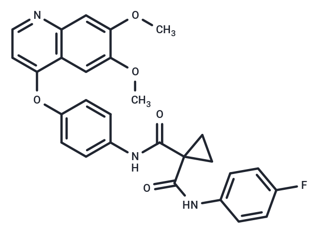
Cabozantinib (XL184) is a multi-targeted tyrosine kinase receptor inhibitor that inhibits VEGFR2, c-Met, Kit, Axl, and Flt3 (IC50=0.035/1.3/4.6/7/11.3 nM). Cabozantinib exhibits both antitumor and antiangiogenic activity.

| Pack Size | Price | Availability | Quantity |
|---|---|---|---|
| 5 mg | $39 | In Stock | |
| 10 mg | $55 | In Stock | |
| 50 mg | $72 | In Stock | |
| 100 mg | $97 | In Stock | |
| 200 mg | $155 | In Stock | |
| 500 mg | $262 | In Stock | |
| 1 mL x 10 mM (in DMSO) | $43 | In Stock |
| Description | Cabozantinib (XL184) is a multi-targeted tyrosine kinase receptor inhibitor that inhibits VEGFR2, c-Met, Kit, Axl, and Flt3 (IC50=0.035/1.3/4.6/7/11.3 nM). Cabozantinib exhibits both antitumor and antiangiogenic activity. |
| Targets&IC50 | VEGFR2:0.035 nM (cell free), RET:5.2 nM (cell free), Axl:7 nM (cell free), MET (Y1248H):3.8 nM (cell free), MET:1.3 nM (cell free), FLT3:11.3 nM (cell free) |
| In vitro | METHODS: Prostate cancer cells LNCaP, C4-2B and PC-3 were treated with Cabozantinib (0.01-5 µM) for 72 h. Cell viability was measured by WST-1 Assay. RESULTS: Cabozantinib inhibited cell viability of LNCaP, C4-2B and PC-3 cell lines in a dose-dependent manner. [1] METHODS: Human renal cancer cells 786-O and A498 were treated with Cabozantinib (10-100 nM) for 1 h, followed by stimulation with HGF (1 nM) for 20 min, and the expression levels of target proteins were detected by Western Blot. RESULTS: 10 nM Cabozantinib treatment inhibited HGF activation of pMET, pAKT, pERK and p-mTOR.[2] |
| In vivo | METHODS: To detect anti-tumor activity in vivo, Cabozantinib (60 mg/kg) was administered orally to SCID mice injected intra-tibially with prostate cancer cells Ace-1 once daily for five weeks. RESULTS: Cabozantinib inhibited the progression of Ace-1 cells in vivo. [1] METHODS: To assay antitumor activity in vivo, Cabozantinib (1-60 mg/kg) was orally administered to nu/nu mice bearing tumors MDA-MB-231, H441, or C6 once daily for 12-14 days. RESULTS: Cabozantinib inhibited tumor growth in a dose-dependent manner. [3] |
| Kinase Assay | The inhibition profile of cabozantinib against a broad panel of 270 human kinases was determined using luciferase-coupled chemiluminescence, 33P-phosphoryl transfer, or AlphaScreen technology. Recombinant human full-length, glutathione S-transferase tag or histidine tag fusion proteins were used, and half maximal inhibitory concentration (IC50) values were determined by measuring phosphorylation of peptide substrate poly(Glu, Tyr) at ATP concentrations at or below the Km for each respective kinase. The mechanism of kinase inhibition was evaluated using the AlphaScreen Assay by determining the IC50 values over a range of ATP concentrations [1]. |
| Cell Research | Receptor phosphorylation of MET, VEGFR2, AXL, FLT3, and KIT were, respectively, assessed in PC3, HUVEC, MDA-MB-231, FLT3-transfected BaF3, and KIT-transfected MDA-MB-231 cells. Cells were serum starved for 3 to 24 hours, then incubated for 1 to 3 hours in serum-free medium with serially diluted cabozantinib before 10-minute stimulation with ligand: HGF (100 ng/mL), VEGF (20 ng/mL), SCF (100 ng/mL), or ANG1 (300 ng/mL). Receptor phosphorylation was determined either by ELISA using specific capture antibodies and quantitation of total phosphotyrosine or immunoprecipitation and Western blotting with specific antibodies and quantitation of total phosphotyrosine. Total protein served as loading controls [1]. |
| Animal Research | Female nu/nu mice were housed according to the Exelixis Institutional Animal Care and Use Committee guidelines. H441 cells (3 × 10^6) were implanted intradermally into the hind flank and when tumors reached approximately 150 mg, tumor weight was calculated using the formula: (tumor volume = length (mm) × width^2 (mm^2)]/2, mice were randomized (n = 5 per group) and orally administered a single 100 mg/kg dose of cabozantinib or vehicle. Tumors were collected at the indicated time points. Pooled tumor lysates were subjected to immunoprecipitation with anti-MET and Western blotting with anti-phosphotyrosine MET. After blot stripping, total MET was quantitated as a loading control. In a separate experiment, naive mice (n = 5 per group) were administered a single 100 mg/kg dose of cabozantinib or vehicle, followed by intravenous administration of HGF (10 μg per mouse) 10 minutes before liver collection. Analysis of MET phosphorylation in liver lysates was as described above. In a separate experiment, naive mice (n = 5 per group) were administered a single 100 mg/kg dose of cabozantinib or vehicle, followed by intravenous administration of VEGF (10 μg per mouse) 30 minutes before lung collection. Pooled lung lysates were subjected to immunoprecipitation with FLK1 and Western blotting with anti-phosphotyrosine. After blot stripping, total FLK1 was quantitated as a loading control [1]. |
| Alias | XL184, BMS-907351 |
| Molecular Weight | 501.51 |
| Formula | C28H24FN3O5 |
| Cas No. | 849217-68-1 |
| Smiles | C(NC1=CC=C(OC=2C3=C(C=C(OC)C(OC)=C3)N=CC2)C=C1)(=O)C4(C(NC5=CC=C(F)C=C5)=O)CC4 |
| Relative Density. | 1.397 g/cm3 |
| Storage | Powder: -20°C for 3 years | In solvent: -80°C for 1 year | Shipping with blue ice. | |||||||||||||||
| Solubility Information | H2O: < 1 mg/mL (insoluble or slightly soluble) DMSO: 50 mg/mL (99.7 mM), Sonication is recommended. 10% DMSO+40% PEG300+5% Tween 80+45% Saline: 9.3 mg/mL (18.54 mM), suspension.In vivo: Please add the solvents sequentially, clarifying the solution as much as possible before adding the next one. Dissolve by heating and/or sonication if necessary. Working solution is recommended to be prepared and used immediately. Ethanol: < 1 mg/mL (insoluble or slightly soluble) | |||||||||||||||
Solution Preparation Table | ||||||||||||||||
DMSO
| ||||||||||||||||

Copyright © 2015-2025 TargetMol Chemicals Inc. All Rights Reserved.