Shopping Cart
- Remove All
 Your shopping cart is currently empty

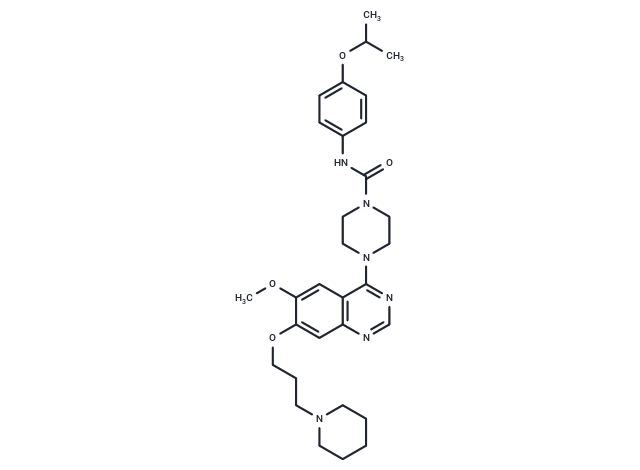
Tandutinib (CT53518) (MLN518, CT53518), an effective FLT3 antagonist (IC50: 0.22 μM), can also inhibit c-Kit and PDGFR, 15-20 fold higher potency for FLT3 versus CSF-1R and >100-fold selectivity for the same target versus FGFR, EGFR, and KDR.

| Pack Size | Price | Availability | Quantity |
|---|---|---|---|
| 25 mg | $38 | In Stock | |
| 50 mg | $54 | In Stock | |
| 100 mg | $73 | In Stock | |
| 200 mg | $102 | In Stock | |
| 1 mL x 10 mM (in DMSO) | $53 | In Stock |
| Description | Tandutinib (CT53518) (MLN518, CT53518), an effective FLT3 antagonist (IC50: 0.22 μM), can also inhibit c-Kit and PDGFR, 15-20 fold higher potency for FLT3 versus CSF-1R and >100-fold selectivity for the same target versus FGFR, EGFR, and KDR. |
| Targets&IC50 | FLT3:0.22 μM |
| In vitro | In mice carrying Ba/F3 cells expressing the W51 FLT3-ITD mutation, Tandutinib (60 mg/kg, twice daily, orally) significantly extends lifespan. In a mouse bone marrow transplantation model, it also significantly reduces mortality rates. Tandutinib (180 mg/kg, twice daily) exhibits minor toxicity to normal hematopoietic function and is effective in treating FLT3 ITD-positive leukemia in mice. |
| In vivo | Unlike Staurosporine, Tandutinib demonstrates superior inhibition of βPDGF, FLT3, and c-Kit autophosphorylation (IC50: 0.17-0.22 μM), while exerting minimal effect on EGFR, FGFR, KDR, InsR, Src, Abl, PKC, PKA, and MAPKs. Tandutinib exhibits specificity towards inhibiting FLT3, effectively inducing apoptosis in FLT3-ITD positive Molm-14 cells (51% and 78% at 24 and 96 hours, respectively) but not in FLT3-ITD negative THP-1 cells. This compound preferentially restricts the explosive colony growth in FLT3 ITD positive AML patients without affecting the colony formation of normal progenitor cells. Moreover, Tandutinib inhibits cell growth independent of IL-3 and suppresses FLT3-ITD autophosphorylation (IC50: 10-100 nM). It also impedes the proliferation of Ba/F3 cells with FLT3-ITD mutation (IC50: 10-30 nM) and targets FLT3-ITD-positive Molm-13 and 14 cells (IC50: 10 nM), showcasing its potential therapeutic value in targeted leukemia treatments. |
| Kinase Assay | Cell based receptor autophosphorylation assays: Autophosphorylation of PDGFR family kinase assays are cell-based enzyme-linked immunosorbent (ELISA) assays using CHO cells expressing wild-type PDGFRβ, chimeric protein PDGFRβ/c-Kit, and PDGFRβ/Flt3 which contain the extracellular and transmembrane domains of PDGFRβ and the cytoplasmic domain of c-Kit, and Flt-3. Cells are grown to confluency in 96-well microtiter plates under standard tissue culture conditions, followed by serum starvation for 16 hours. Briefly, quiescent cells are incubated at 37 °C with increasing concentrations of Tandutinib for 30 minutes followed by the addition of 8 nM PDGF-BB for 10 minutes. Cells are lysed in 100 mM Tris, pH 7.5, 750 mM NaCl, 0.5% Triton X-100, 10 mM sodium pyrophosphate, 50 mM NaF, 10 μg/mL aprotinin, 10 μg/mL leupeptin, 1 mM phenylmethylsulfonyl fluoride, 1 mM sodium vanadate, and the lysate is cleared by centrifugation at 15,000 g for 5 minutes. Clarified lysates are transferred into a second microtiter plate in which the wells are previously coated with 500 ng/well of 1B5B11 anti-PDGFRβ mAb and then incubated for 2 hours at room temperature. After washing three times with binding buffer (0.3% gelatin, 25 mM HEPES, pH 7.5, 100 mM NaCl, 0.01% Tween 20), 250 ng/mL of rabbit polyclonal anti-phosphotyrosine antibody is added and plates are incubated at 37 °C for 60 minutes. Subsequently, each well is washed three times with binding buffer and incubated with 1 μg/mL of horseradish peroxidase-conjugated anti-rabbit antibody at 37 °C for 60 minutes. Wells are washed prior to adding 2,2'-azino-bis(3-ethylbenzthiazoline-6-sulfonic acid), and the rate of substrate formation is monitored at 650 nm. |
| Cell Research | Cells are exposed to increasing concentrations of Tandutinib (0.004-30 μM). Cells are grown for 3-7 days in tissue culture, and viable cells, determined by Trypan blue dye exclusion, are counted. At daily intervals, cells are harvested, washed, and resuspended in 100 uL binding buffer containing 10 mM HEPES (pH 7.4), 140 mM NaCl, and 2.5 mM CaCl2. Annexin V-FITC (100 ng) and propidium iodide (250 ng) are added to the cell suspension followed by incubation at room temperature for 15 minutes. Flow cytometry is performed immediately after staining on a FACSort flow cytometer with excitation at 488 nm. Fluorescence of annexin V-FITC and DNA propidium iodide staining are measured at 515 nm and 585 nm, respectively.(Only for Reference) |
| Alias | NSC726292, MLN518, CT53518 |
| Molecular Weight | 562.7 |
| Formula | C31H42N6O4 |
| Cas No. | 387867-13-2 |
| Smiles | O(C)C1=CC=2C(=NC=NC2C=C1OCCCN3CCCCC3)N4CCN(C(NC5=CC=C(OC(C)C)C=C5)=O)CC4 |
| Relative Density. | 1.213 g/cm3 |
| Storage | Powder: -20°C for 3 years | In solvent: -80°C for 1 year | Shipping with blue ice. | |||||||||||||||||||||||||||||||||||
| Solubility Information | Ethanol: 6 mg/mL (10.66 mM), Sonication is recommended. DMSO: 50 mg/mL (88.86 mM), Sonication is recommended. | |||||||||||||||||||||||||||||||||||
Solution Preparation Table | ||||||||||||||||||||||||||||||||||||
Ethanol/DMSO
DMSO
| ||||||||||||||||||||||||||||||||||||

Copyright © 2015-2025 TargetMol Chemicals Inc. All Rights Reserved.