Shopping Cart
- Remove All
 Your shopping cart is currently empty

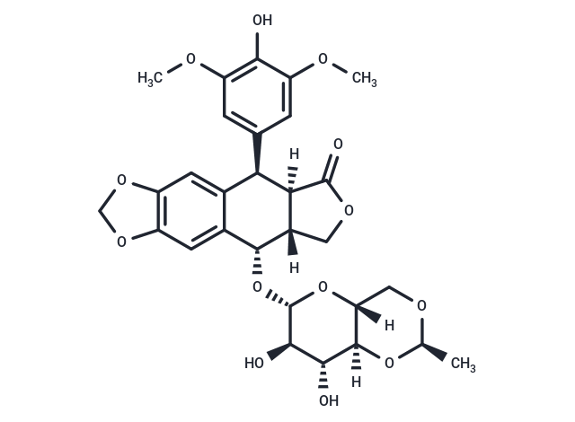
Etoposide (VP-16-213) is a topoisomerase II inhibitor that inhibits DNA synthesis by forming a complex with topoisomerase II and DNA (IC50=60.3 μM). Etoposide has antitumor activity and induces apoptosis and autophagy.

| Pack Size | Price | Availability | Quantity |
|---|---|---|---|
| 50 mg | $33 | In Stock | |
| 100 mg | $48 | In Stock | |
| 500 mg | $108 | In Stock | |
| 1 mL x 10 mM (in DMSO) | $50 | In Stock |
| Description | Etoposide (VP-16-213) is a topoisomerase II inhibitor that inhibits DNA synthesis by forming a complex with topoisomerase II and DNA (IC50=60.3 μM). Etoposide has antitumor activity and induces apoptosis and autophagy. |
| Targets&IC50 | Topo II:60.3 μM |
| In vitro | METHODS: Human cervical cancer cells HeLa were treated with Etoposide (25-400 μM) for 24-48 h, and cell viability was measured by MTT. RESULTS: Etoposide inhibited the proliferation of Hela cells with IC50s of 167.3 μM and 52.7 μM for 24 h and 48 h, respectively. The IC50 of Etoposide was 167.3 μM and 52.7 μM at 24 h and 48 h, respectively. [1] METHODS: Human lung adenocarcinoma cells A549 were treated with Etoposide (0.75-3 μM) for 4 h. The cell cycle was detected by Flow Cytometry. RESULTS: Etoposide caused a significant decrease in the percentage of A549 cells in G0/G1 and S phases. Meanwhile, the percentage of A549 cells in G2/M phase was significantly increased. [2] METHODS: Mouse embryonic fibroblast MEFs were treated with Etoposide (1.5-150 μM) for 3-18 h, and the expression levels of target proteins were detected by Western Blot. RESULTS: 150 μM Etoposide induced a strong cleavage of caspase-3 within 6 h, while 1.5 or 15 μM activated caspase-3 only after 18 h. [3] |
| In vivo | METHODS: To detect anti-tumor activity in vivo, Etoposide (10 mg/kg) and Cisplatin (5-7.5 mg/kg) were intraperitoneally injected every two days for two weeks into KSN nude mice harboring human endometrial adenocarcinoma tumors Ishikawa. RESULTS: Etoposide, as a single agent, had little or no inhibitory effect on tumor growth, while the combination of Etoposide and Cisplatin significantly inhibited tumor growth. [4] METHODS: To detect anti-tumor activity in vivo, Etoposide (80 mg/kg in 0.5% methylcellulose) was administered by gavage to immunodeficient mice harboring human glioblastoma tumor U87 once a day for 40 days. RESULTS: 80 mg/kg Etoposide inhibited U87 tumor growth by 95%. [5] |
| Kinase Assay | Nuclear extracts are prepared, and nuclei are isolated. The activity of topoisomerase II is calculated from the percentage of decatenation obtained. Tritiated kinoplast DNA (KDNA 0.22 μg) is used as a substrate. Etoposide and topoisomerase II are incubated for 30 min at 37 ℃ and are stopped with 1% sodium dodecyl sulfate (SDS) and proteinase K (100 μg/mL). The percentages of decatenation and inhibition of topoisomerase II by Etoposide are obtained [5]. |
| Cell Research | After the Etoposide treatment, cells are removed from the dish with phosphate-buffered saline (PBS) containing 0.03% trypsin and 0.27 mM ethylenediaminetetraacetic acid (EDTA) and are diluted into culture dishes in appropriate numbers to yield between 20 and 200 colonies. After 12 days, cultures are fixed with methanol-acetic acid, stained with crystal violet, and scored for colonies containing more than 50 cells [5]. |
| Animal Research | The in vivo model for nude mice HB (NMHB) has been established. Only HB cells with embryonal components are grafted and reproduced successfully in this model. Each NMHB subsequently is transplanted into 50 mice for treatment groups. Treatment is initiated when the majority of the tumors reach a volume of 50-100 mm3. The mice are stratified according to their tumor volume and randomly assigned to groups of ten animals each. The animals injected with tumor are given ifosfamide, cisplatin, doxorubicin, etoposide (10 mg/kg/day, i.v.), and carboplatin as single agents in two blocks. One group of ten animals for each original xenograft served as a control group. After initiation of treatment, the tumor growth is recorded at 5-day intervals for 25-30 days and the relative tumor volumes are calculated. Twenty-four hours before the animals are sacrificed, bromodeoxyuridine (BrdU) is injected intraperitoneally for the semiquantitative determination of proliferation activity of the tumor cells (50 μg of BrdU/g body weight) [4]. |
| Alias | VP-16-213, VP-16 |
| Molecular Weight | 588.56 |
| Formula | C29H32O13 |
| Cas No. | 33419-42-0 |
| Smiles | O=C1[C@@]2([C@@H](C=3C([C@@H](O[C@@H]4O[C@]5([C@]([C@H](O)[C@H]4O)(O[C@H](C)OC5)[H])[H])[C@]2(CO1)[H])=CC6=C(C3)OCO6)C7=CC(OC)=C(O)C(OC)=C7)[H] |
| Relative Density. | 1.55 g/cm3 |
| Storage | keep away from direct sunlight | Powder: -20°C for 3 years | In solvent: -80°C for 1 year | Shipping with blue ice. | ||||||||||||||||||||
| Solubility Information | 10% DMSO+40% PEG300+5% Tween 80+45% Saline: 5.89 mg/mL (10.01 mM), In vivo: Please add the solvents sequentially, clarifying the solution as much as possible before adding the next one. Dissolve by heating and/or sonication if necessary. Working solution is recommended to be prepared and used immediately. DMSO: 60 mg/mL (101.94 mM), Sonication is recommended. | ||||||||||||||||||||
Solution Preparation Table | |||||||||||||||||||||
DMSO
| |||||||||||||||||||||

Copyright © 2015-2025 TargetMol Chemicals Inc. All Rights Reserved.